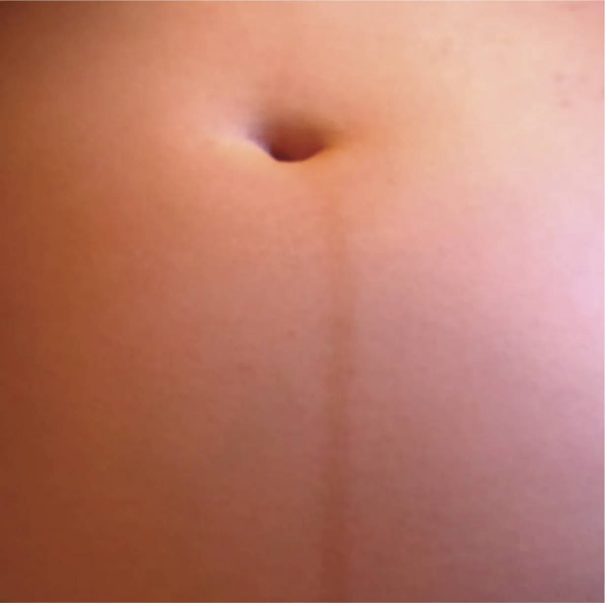
The appearance of the linea nigra, a distinct dark line running down the center of the abdomen, is a common and fascinating physiological change observed during pregnancy. This image clearly depicts the linea nigra on an abdomen at 22 weeks gestation, extending vertically from the umbilicus towards the pubis. While it may be a cause for curiosity or concern for some, it is a benign dermatological manifestation of the profound hormonal shifts occurring during pregnancy. This article will explore the characteristics, causes, and temporary nature of this unique pregnancy marker.
Visible Features of the Linea Nigra
While the image does not contain explicit labels, it clearly illustrates the primary features of the linea nigra.
Linea Nigra: This is the dark, brownish line visible in the center of the abdomen, extending downwards from the navel. Its prominence can vary among individuals, influenced by skin tone and hormonal levels.
Umbilicus (Navel): The central point from which the linea nigra originates superiorly (though it often extends above the navel as well, becoming more prominent below). In the image, the umbilicus is clearly visible as an indented area.
Abdomen: The surrounding area of the skin, which in pregnancy will be stretching to accommodate the growing uterus. The skin tone and texture around the linea nigra are also part of the overall assessment of pregnancy-related skin changes.
The Physiology Behind the Linea Nigra
The linea nigra is a darkened pigmentation of the linea alba, a fibrous band that runs vertically along the midline of the abdomen, formed by the fusion of the aponeuroses of the abdominal muscles. This line is present in everyone, but it is typically pale and barely visible. During pregnancy, however, it becomes hyperpigmented due to hormonal changes, particularly increased levels of estrogen and melanocyte-stimulating hormone (MSH). These hormones stimulate melanocytes, the cells responsible for producing melanin, the pigment that gives skin its color.
The increased melanin production is not confined to the linea alba; it also accounts for other common pregnancy-related hyperpigmentation, such as darkening of the nipples and areolae, and melasma (chloasma) or the “mask of pregnancy” on the face. While more pronounced in individuals with darker skin tones, the linea nigra can appear in women of all ethnicities. Its appearance usually becomes noticeable around the second trimester and darkens progressively throughout the pregnancy.
- The linea nigra is entirely harmless and poses no medical risk to either the mother or the fetus.
Following childbirth, the hormonal levels gradually return to their pre-pregnancy state. As a result, the melanocytes reduce their melanin production, and the linea nigra typically fades over a few weeks or months. In some individuals, a faint trace may persist indefinitely, but it generally becomes much less noticeable.
Conclusion
The linea nigra is a classic example of the fascinating dermatological changes that can occur during pregnancy, driven by profound hormonal shifts. This image at 22 weeks gestation provides a clear visual of this dark medial line, a benign and temporary marker of the pregnant state. While its appearance is a normal physiological response, understanding its etiology can alleviate any concerns and highlight the intricate interplay of hormones in shaping the maternal body throughout gestation.

