Magnetic Resonance Angiography (MRA) stands as a cornerstone of modern diagnostic neuroradiology, providing a non-invasive window into the complex vascular architecture that supplies the human brain. The provided image illustrates a coronal Maximum Intensity Projection (MIP) of the head and neck vessels, tracing the journey of blood from the aortic arch through the cervical region and into the intracranial space. This modality is essential for clinicians to evaluate blood flow, identify structural abnormalities, and plan surgical or endovascular interventions without the risks associated with ionizing radiation or arterial catheterization.
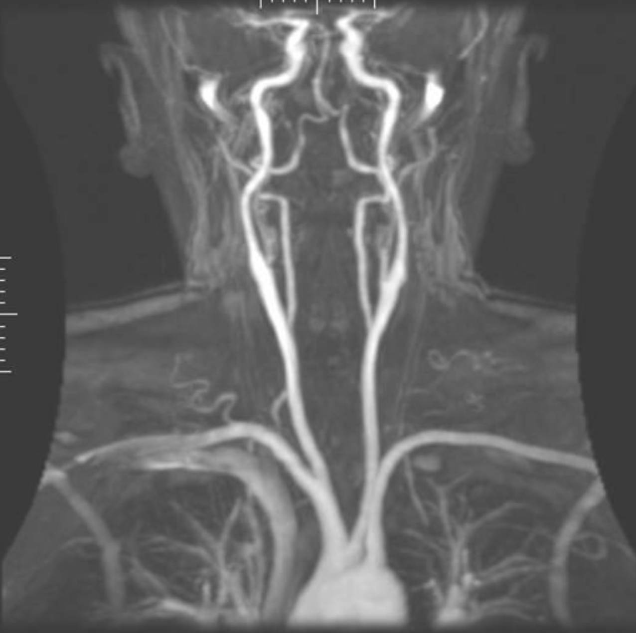
Image Overview
The image presents a comprehensive view of the major arteries of the neck and head. At the base of the image, the superior portion of the aortic arch is visible, serving as the origin for the primary supra-aortic trunks. We can clearly see the brachiocephalic trunk (innominate artery) on the patient’s right side, which subsequently bifurcates into the right common carotid artery and the right subclavian artery. On the left side, the left common carotid artery and the left subclavian artery arise directly from the aortic arch.
Moving superiorly, the bilateral common carotid arteries ascend through the neck. At approximately the level of the upper thyroid cartilage (C3-C4 level), the bifurcation into the internal and external carotid arteries is evident. The internal carotid arteries (ICA) can be traced as they enter the skull base to supply the anterior cerebral circulation. Parallel to the carotids, the vertebral arteries are visible arising from the subclavian arteries, ascending through the transverse foramina of the cervical vertebrae to eventually join and form the basilar artery, providing the posterior circulation.
Anatomical Overview
The cerebrovascular system is divided into the anterior and posterior circulations, both of which are beautifully highlighted in this MRA. The anterior circulation is dominated by the carotid system. The Internal Carotid Artery is particularly vital as it lacks branches in the neck, preserving its high-pressure flow until it reaches the carotid canal of the temporal bone. Once intracranial, it contributes significantly to the Circle of Willis.
The posterior circulation, or the vertebrobasilar system, begins with the vertebral arteries. These vessels are unique because of their protected path within the bony cervical spine. Any compromise in these vessels, such as dissection or atherosclerotic plaque, can lead to posterior circulation strokes, affecting the cerebellum and brainstem. The visual continuity seen in this image allows radiologists to assess for segments of narrowing (stenosis) or pathological dilations (aneurysms) along the entire course of these vessels.
Functional Significance of MRA Techniques
The clarity of this image is achieved through specific MRI sequences and post-processing techniques. Most MRAs of the head and neck utilize either Time-of-Flight (TOF) or Contrast-Enhanced (CE) methods. TOF MRA relies on the movement of blood into a slice to create contrast, making it ideal for intracranial imaging where the use of gadolinium might be avoided. CE-MRA, often used for the neck, utilizes intravenous contrast to shorten the T1 relaxation time of blood, allowing for high-resolution imaging of the carotid and vertebral origins.
The image shown is a Maximum Intensity Projection (MIP). This is a volume-rendering technique that extracts the brightest pixels (representing high-velocity blood flow or contrast) from a stack of 2D slices and projects them into a single 3D-like frame. While MIPs provide an excellent anatomical overview, clinicians must be aware that they can occasionally overestimate the severity of a stenosis because signal loss can occur in areas of highly turbulent flow.
Clinical and Diagnostic Importance
In clinical practice, this type of imaging is the first line of defense when a patient presents with symptoms of a Transient Ischemic Attack (TIA) or stroke. It allows for the rapid identification of carotid artery stenosis, a major risk factor for ischemic events. Beyond atherosclerosis, MRA is superior at detecting arterial dissections—tears in the inner lining of the artery wall—which are common causes of stroke in younger populations.
Furthermore, the visualization of the entire vascular tree helps in diagnosing subclavian steal syndrome, where a blockage in the subclavian artery causes blood to flow backward down the vertebral artery to supply the arm, “stealing” blood from the brain. By mapping these pathways, healthcare providers can determine if a patient is a candidate for carotid endarterectomy, stenting, or medical management to stabilize hemodynamics and prevent future neurological deficits.
Educational Value for Medical Learners
For students and healthcare professionals, this image serves as a vital tool for correlating textbook anatomy with clinical radiology. Understanding the branching patterns of the aortic arch is fundamental; for instance, recognizing that the left common carotid arises directly from the arch while the right arises from the brachiocephalic trunk is a classic anatomical point frequently tested and clinically relevant during catheterization. Observing the spatial relationship between the carotid and vertebral systems helps learners visualize how collateral circulation via the Circle of Willis might compensate for a proximal arterial blockage.
In summary, the MRA of the cerebrovascular highway is more than just a picture; it is a dynamic map of life-sustaining flow. Mastery of this image allows for better diagnostic accuracy and a deeper understanding of the vascular pathologies that threaten brain health.
Medical Learning Tips
- Identify the 'bovine arch' variant where the left common carotid artery shares a common origin with the brachiocephalic trunk.
- Remember that the internal carotid artery typically has no branches in the cervical region, distinguishing it from the external carotid.
- Be cautious with MIP images, as they can exaggerate the degree of vascular stenosis in regions of turbulent flow.

