Discover the vital journey of blood circulation within the human body, a continuous process where blood flows through the heart, lungs, and various organs and tissues. This detailed explanation clarifies how deoxygenated blood becomes oxygenated in the lungs before being distributed, eventually returning to the heart. Grasp the fundamental mechanisms that ensure every cell receives the oxygen and nutrients it needs for life.
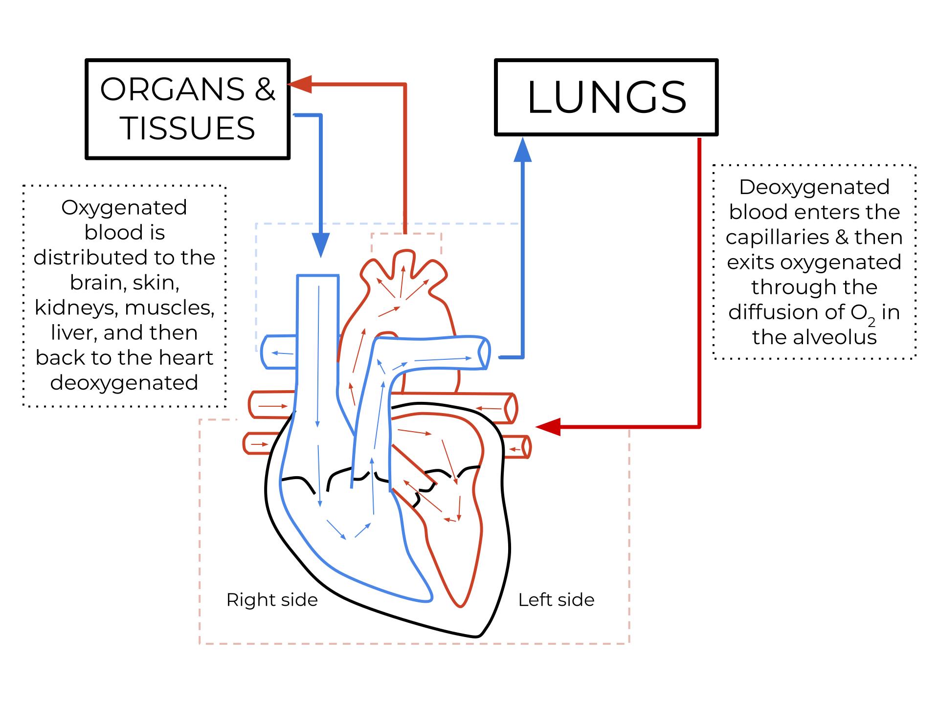
ORGANS & TISSUES: This label represents all the body’s functional units, apart from the lungs, that require oxygenated blood to perform their metabolic processes. These include the brain, skin, kidneys, muscles, and liver, which receive blood from the heart’s left side.
LUNGS: The lungs are the primary organs responsible for gas exchange, where deoxygenated blood releases carbon dioxide and picks up oxygen. This vital process occurs within tiny air sacs called alveoli, ensuring the blood is re-oxygenated before returning to the heart.
Oxygenated blood is distributed to the brain, skin, kidneys, muscles, liver, and then back to the heart deoxygenated: This descriptive text explains the systemic circulation pathway. Oxygen-rich blood from the left side of the heart is pumped to various body parts, delivering oxygen and nutrients, and then returns to the right side of the heart as deoxygenated blood.
Deoxygenated blood enters the capillaries & then exits oxygenated through the diffusion of O2 in the alveolus: This label describes the pulmonary circulation and the process of gas exchange in the lungs. Deoxygenated blood travels to the lungs, where it enters tiny capillaries surrounding the alveoli, picks up oxygen via diffusion, and then becomes oxygenated.
Right side: This refers to the right atrium and right ventricle of the heart, which handle deoxygenated blood. The right side receives deoxygenated blood from the body and pumps it to the lungs.
Left side: This refers to the left atrium and left ventricle of the heart, which handle oxygenated blood. The left side receives oxygenated blood from the lungs and pumps it to the rest of the body.
The human circulatory system is an incredibly efficient and complex network designed to transport essential substances throughout the body. At its core is the heart, a powerful muscular pump that drives blood through two interconnected circuits: the pulmonary circulation and the systemic circulation. This diagram beautifully illustrates the elegant simplicity and crucial interdependence of these two pathways, highlighting how deoxygenated blood is revitalized with oxygen in the lungs before being distributed to every cell and tissue.
Understanding the continuous loop of blood circulation is fundamental to comprehending human physiology. Blood, a vital fluid, acts as the body’s transportation system, carrying oxygen, nutrients, hormones, and immune cells to where they are needed, while simultaneously collecting metabolic waste products like carbon dioxide. The heart’s four chambers work in perfect synchrony to prevent the mixing of oxygenated and deoxygenated blood, ensuring maximum efficiency in delivering life-sustaining resources.
The journey begins with deoxygenated blood entering the right side of the heart, destined for the lungs. Once oxygenated, this enriched blood returns to the left side of the heart, ready to be propelled into the vast network of arteries and capillaries that reach every corner of the body. This rhythmic cycle, powered by the heart’s relentless beat, is crucial for maintaining cellular function, organ integrity, and overall well-being. Any disruption in this delicate balance can have significant health implications, underscoring the importance of a healthy cardiovascular system.
- The circulatory system has two main circuits: pulmonary and systemic.
- The heart acts as a central pump for blood.
- Lungs are responsible for oxygenating blood.
- Organs and tissues receive oxygen and nutrients from blood.
- Deoxygenated and oxygenated blood are kept separate in the heart.
The Pulmonary Circuit: Recharging with Oxygen
The journey of deoxygenated blood begins as it returns from the body’s organs and tissues, laden with carbon dioxide, and enters the right side of the heart. Specifically, it first flows into the right atrium, then moves into the right ventricle. From the right ventricle, this deoxygenated blood is pumped into the pulmonary artery, which branches to carry it to both lungs. Within the lungs, the pulmonary arteries divide into a vast network of tiny capillaries that surround the alveoli, the microscopic air sacs where gas exchange takes place. Here, through the process of diffusion, carbon dioxide is released from the blood into the alveoli to be exhaled, and crucially, oxygen from the inhaled air diffuses from the alveoli into the blood. Once oxygenated, this now oxygen-rich blood collects in the pulmonary veins, which then carry it back to the left atrium of the heart, completing the pulmonary circuit. This critical step ensures that the blood is fully revitalized with oxygen, ready for its next mission.
The Systemic Circuit: Delivering Life to Every Cell
Upon returning to the left atrium from the lungs, the freshly oxygenated blood then flows into the left ventricle, the most muscular chamber of the heart. The left ventricle contracts powerfully to pump this oxygenated blood into the aorta, the body’s largest artery. From the aorta, the blood embarks on its journey through the systemic circuit, branching into numerous arteries that progressively diminish in size, eventually becoming arterioles and then capillaries. It is within these microscopic capillaries that the essential exchange occurs: oxygen and nutrients are delivered to the cells of organs and tissues (such as the brain, skin, kidneys, muscles, and liver), while carbon dioxide and other waste products are picked up from these cells. After this vital exchange, the now deoxygenated blood, rich in carbon dioxide, begins its return journey through venules, which merge to form larger veins. These veins ultimately converge into the superior and inferior vena cavae, which empty into the right atrium of the heart, restarting the entire cycle.
The continuous and efficient operation of blood circulation is fundamental to maintaining life and health. Each component, from the powerful pump of the heart to the delicate exchange sites in the lungs and capillaries, plays an indispensable role. Understanding this intricate interplay between the pulmonary and systemic circuits provides crucial insight into how the body sustains itself, and highlights the importance of maintaining cardiovascular health through lifestyle choices and medical care

