In the vast kingdom of Fungi, the ability to transition between different structural forms is one of the most significant evolutionary adaptations. While yeasts are characterized by their unicellular nature, many fungi exist as multicellular organisms known as molds. The fundamental structural unit of these molds is the hypha, a long, branching filamentous structure that collectively forms a network called a mycelium. In a clinical and microbiological context, the microscopic appearance of these filaments is a primary diagnostic tool used to differentiate between various fungal pathogens. The presence or absence of cross-walls, the pattern of branching, and the diameter of the filaments can provide critical clues regarding the identity of an infecting agent. Understanding Fungal morphology is not merely an academic exercise; it is the cornerstone of laboratory mycology, allowing clinicians to distinguish between relatively benign environmental contaminants and highly aggressive, life-threatening pathogens. This article explores the three primary types of hyphal structures—septate, coenocytic (nonseptate), and pseudohyphae—and examines their physiological significance, diagnostic characteristics, and clinical implications in human health.
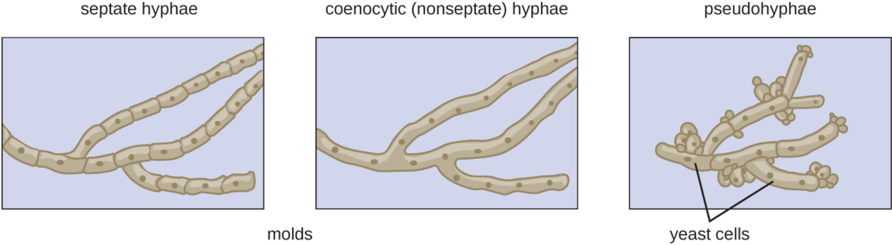
septate hyphae: These are filamentous structures divided into individual cells by internal cross-walls known as septa. While these septa provide structural reinforcement, they typically contain microscopic pores that allow for the regulated movement of cytoplasm, organelles, and nutrients between adjacent cells.
coenocytic (nonseptate) hyphae: These hyphae lack internal cross-walls, resulting in long, continuous multinucleated tubes where the cytoplasm flows freely throughout the entire structure. This architecture is characteristic of fungi in the Zygomycota phylum and facilitates extremely rapid growth and nutrient transport.
pseudohyphae: These consist of chains of elongated yeast cells that remain attached after the process of budding, creating a structure that resembles true hyphae but lacks structural continuity. They are a hallmark of opportunistic pathogens like Candida albicans and represent a specialized growth phase designed for tissue invasion.
molds: This label refers to the multicellular, filamentous growth form of fungi depicted in the first two diagrams. Molds use their hyphal networks to colonize substrates, secrete digestive enzymes, and produce reproductive spores known as conidia.
yeast cells: These are the unicellular forms of fungi that typically reproduce by an asymmetrical division process called budding. When these cells fail to detach during rapid growth or under specific environmental triggers, they form the pseudohyphal chains shown in the third panel.
Classification of Fungal Morphology in Microbiology
The study of Mycology recognizes that fungi are remarkably plastic in their growth forms. Most fungi can be categorized based on whether they grow as yeasts (single cells) or molds (multicellular filaments). However, many significant human pathogens are dimorphic, meaning they can switch between these two forms depending on environmental factors such as temperature, pH, and nutrient availability. For instance, many systemic pathogens like Histoplasma capsulatum exist as molds in the cold environment of the soil but transform into yeasts at the 37°C temperature of the human body.
Hyphae are the engine of mold growth. They grow exclusively at their tips through a highly polarized process involving the transport of vesicles to the apex. This growth pattern allows fungi to explore their environment and penetrate solid surfaces, including human tissue. The internal organization of these hyphae—whether they are segmented by septa or exist as a continuous coenocytic mass—determines how the fungus responds to damage and how quickly it can distribute resources. In a laboratory setting, identifying these structures via microscopic examination of a KOH prep or a GMS stain is often the first step in directing patient care.
Septate Hyphae: Precision and Protection
Septate hyphae are found in the most common and clinically significant fungi, including members of the Ascomycota and Basidiomycota phyla. The defining feature, the septum, is a complex biological structure. In many species, these septa are incomplete, meaning they have a central pore. This pore allows the fungus to function as a single integrated unit while still maintaining cellular boundaries. Specialized organelles called Woronin bodies are often located near these pores; if a hyphal segment is damaged, these bodies can quickly plug the pore to prevent the loss of cytoplasm from the rest of the filament. This “cellular sealing” mechanism makes septate fungi highly resilient to physical trauma.
Common examples of septate fungi encountered in medical settings include Aspergillus and Penicillium species, as well as the dermatophytes responsible for skin infections like athlete\’s foot. Under the microscope, septate hyphae usually appear as thin, uniform filaments (typically 3–5 micrometers in diameter) with distinct cross-walls appearing at regular intervals. They often branch at acute, 45-degree angles. This predictable and organized structure is a key diagnostic feature that helps pathologists rule out more primitive, nonseptate molds during tissue biopsy analysis.
Coenocytic Hyphae: The Non-segmented Powerhouse
Coenocytic hyphae, also known as nonseptate or pauciseptate hyphae, represent a more primitive but highly efficient evolutionary strategy. These filaments lack regular cross-walls, essentially forming a massive, multi-nucleated super-cell. Because there are no internal barriers, the internal transport of proteins and organelles is extremely rapid, allowing these fungi to grow much faster than their septate counterparts. This rapid growth is a survival mechanism that allows them to quickly overwhelm a substrate—or a host.
From a clinical perspective, coenocytic hyphae are most famously associated with the Mucormycetes (formerly Zygomycetes), the agents of mucormycosis. These fungi are notorious for causing aggressive, angioinvasive infections in immunocompromised patients, particularly those with uncontrolled diabetes. Microscopically, coenocytic hyphae are distinct from septate ones: they are much wider (often 10–20 micrometers), appear ribbon-like and irregular, and branch at wide, 90-degree angles. Because they lack the structural reinforcement of septa, they are fragile and often appear twisted or collapsed in histological sections, a feature that provides a vital clue for rapid diagnosis and emergency treatment.
Pseudohyphae: The Invasive Phase of Yeast
Pseudohyphae represent a unique middle ground between unicellular yeast and true multicellular molds. Unlike true hyphae, which grow from a germ tube and have a continuous cell wall, pseudohyphae are formed by the successive budding of yeast cells that fail to separate. This results in a chain of elongated cells with distinct “constrictions” at the sites of attachment, resembling a string of sausages. This phenotypic switch is a critical component of the virulence of Candida albicans, the most common fungal pathogen in humans.
The transition to pseudohyphal and true hyphal growth in yeast is often triggered by the conditions found within a host, such as body temperature and the presence of serum. This change in shape is more than just cosmetic; it allows the yeast to escape immune cells like macrophages and physically push through epithelial and endothelial layers to enter the bloodstream. In a clinical lab, finding pseudohyphae in a vaginal swab or a sputum sample is often indicative of an active infection rather than simple colonization. The presence of these structures signifies that the yeast has switched into its invasive, pathogenic mode.
Laboratory Diagnostics and Microscopy Techniques
The definitive identification of fungal structures relies on specialized staining and preparation techniques. Because fungal cell walls are composed of chitin and glucans, they do not stain well with standard Gram stains. Instead, clinicians use a KOH (potassium hydroxide) prep to dissolve human tissue and debris, leaving the resilient fungal walls clearly visible. For tissue biopsies, Gomori Methenamine Silver (GMS) and Periodic Acid-Schiff (PAS) stains are the gold standards, highlighting the hyphal structure against a contrasting background.
When a microbiologist looks at these slides, they are performing a process of elimination based on the diagrams shown above. If the hyphae are thin, regularly septate, and branch at acute angles, Aspergillus is a high possibility. If the hyphae are broad, ribbon-like, and lack septa, Mucor or Rhizopus is suspected. If there are oval yeast cells mixed with chains of elongated cells with constrictions, Candida is the likely culprit. This morphological assessment is often much faster than waiting for a fungal culture to grow, which can take several days or even weeks.
Pathogenesis and Antifungal Therapy Implications
The structural type of the invading fungus has direct consequences for the patient’s prognosis and the choice of Antifungal therapy. Septate molds like Aspergillus often cause chronic infections or allergic responses, although they can become invasive in severely neutropenic patients. In contrast, nonseptate molds are characterized by their ability to invade blood vessels (angioinvasion), leading to tissue infarction and necrosis. Because nonseptate fungi grow so rapidly and cause so much damage so quickly, they require aggressive surgical debridement in addition to high-dose antifungal medication.
Most modern antifungals, such as azoles and echinocandins, target the fungal cell wall or the synthesis of ergosterol in the cell membrane. While these drugs are effective across various morphologies, the resistance patterns can vary. For example, Candida species that are forming extensive pseudohyphae and biofilms may be more resistant to treatment than their unicellular yeast forms. Understanding the hyphal structure helps clinicians predict how a fungus might spread and which medications will be most effective at penetrating the fungal mass to reach the living cells within.
Conclusion: The Structural Foundation of Mycology
The complexity of fungal life is perfectly illustrated by the diversity of its hyphal forms. From the reinforced, segmented pipes of septate hyphae to the high-speed cytoplasmic highways of coenocytic tubes and the invasive chains of pseudohyphae, each structure represents a specialized tool for survival and infection. For the medical community, these microscopic differences are the primary language of fungal identification. By mastering the visual characteristics of these filaments, researchers and clinicians can unlock the mysteries of fungal pathogenesis and provide targeted, life-saving interventions. As we face a rising incidence of opportunistic fungal infections in an increasingly vulnerable global population, the fundamental knowledge of hyphal morphology remains our most reliable first line of defense. The ability to see and understand the “little rooms” and “long tubes” of the fungal world is, ultimately, what allows us to protect the human body from the quiet, persistent growth of these ancient organisms.
Fungal Hyphae, Septate, Coenocytic, Pseudohyphae, Microbiology, Mycology, Candida, Aspergillus, Mold, Yeast

