Understand the critical symptoms of a heart attack, a medical emergency caused by a blocked coronary artery. This article explains the sensation of tightness or pain in the chest, a hallmark sign of myocardial infarction, and the underlying physiological event. Learn to identify these vital indicators to ensure prompt medical attention and improve outcomes during a cardiac event.
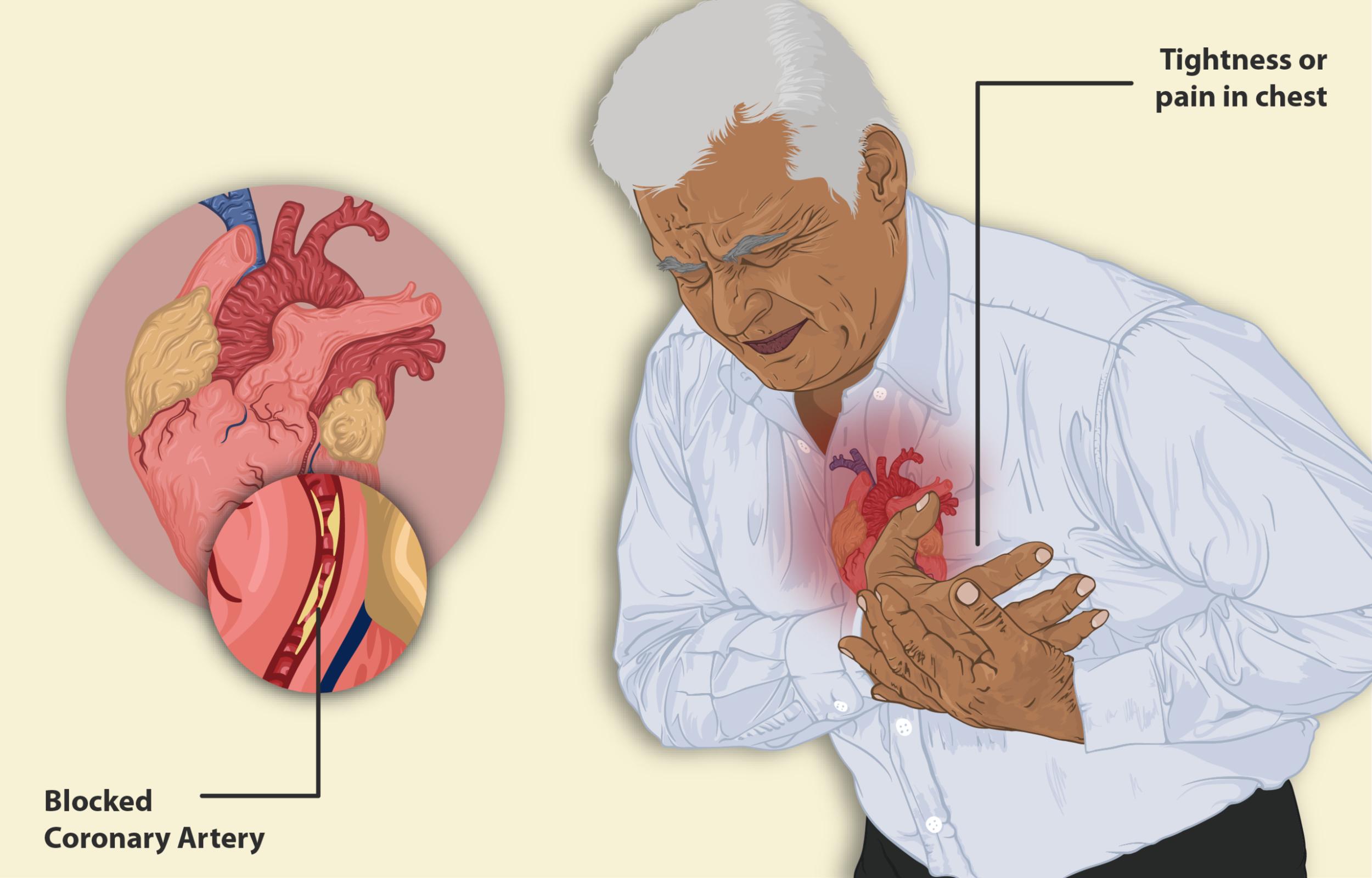
Tightness or pain in chest: This refers to the primary symptom experienced during angina or a heart attack, often described as a crushing, squeezing, or heavy sensation. This discomfort can vary in intensity and may radiate to other parts of the body, indicating the heart muscle is not receiving sufficient oxygen.
Blocked Coronary Artery: This label highlights the direct cause of a heart attack, where one of the arteries supplying blood to the heart muscle becomes severely narrowed or completely obstructed. This blockage, typically due to a rupture of an atherosclerotic plaque and subsequent clot formation, prevents oxygen-rich blood from reaching a section of the heart muscle, leading to tissue damage.
A heart attack, medically termed myocardial infarction (MI), is a critical medical emergency that occurs when blood flow to a part of the heart muscle is severely reduced or completely blocked. This deprivation of oxygen and nutrients causes damage or death to the affected heart tissue. The most common cause is the buildup of plaque in the coronary arteries, a condition known as atherosclerosis. When this plaque ruptures, a blood clot can form rapidly, obstructing the artery and initiating a heart attack. Recognizing the symptoms promptly is vital, as immediate medical attention can significantly limit heart damage and save lives. The illustration vividly depicts a man experiencing the characteristic chest discomfort associated with such an event, alongside a detailed view of the obstructed artery.
The Mechanism of a Heart Attack
Atherosclerosis is a chronic disease where fatty deposits, cholesterol, calcium, and other substances accumulate in the inner lining of the arteries, forming plaques. These plaques can grow over time, narrowing the arteries and reducing blood flow. The danger escalates when a plaque ruptures. When this happens, the body’s clotting mechanism is activated, leading to the formation of a blood clot (thrombus) at the site of the rupture. If this clot is large enough, it can completely block the coronary artery, cutting off the blood supply to the part of the heart muscle downstream from the blockage. This sudden lack of oxygen, known as ischemia, causes the heart muscle cells to begin to die, leading to a myocardial infarction.
Symptoms of a Heart Attack
While chest pain or tightness is the most recognized symptom, heart attack symptoms can vary significantly among individuals, and some people, especially women, older adults, and those with diabetes, may experience more subtle signs.
Common symptoms include:
- Chest discomfort: This can be felt as pressure, squeezing, fullness, or pain in the center of the chest that lasts more than a few minutes, or goes away and comes back.
- Pain radiating to other areas: Discomfort can spread to one or both arms (especially the left arm), the back, neck, jaw, or stomach.
- Shortness of breath: This can occur with or without chest discomfort.
- Other signs: Cold sweat, nausea, lightheadedness, or sudden dizziness.
It’s crucial to understand that not everyone experiencing a heart attack will have all these symptoms, and the intensity can vary. Any new, unexplained, or persistent chest discomfort or other related symptoms should be considered a medical emergency.
In conclusion, a heart attack is a serious medical event caused by the blockage of a coronary artery, leading to a critical reduction in blood flow to the heart muscle. Recognizing the warning signs, particularly tightness or pain in the chest, and understanding that these symptoms can spread to other parts of the body, is paramount for a rapid response. Timely medical intervention, ideally within the first few hours of symptom onset, is crucial for restoring blood flow to the heart, minimizing damage, and significantly improving the chances of survival and recovery.

