The cerebellum, a vital structure for coordination and balance, relies on intricate neural pathways to communicate with the rest of the brain and spinal cord. This diagram showcases the cerebellar peduncles, the three key bundles of nerve fibers—superior, middle, and inferior—that serve as the primary connections, each originating from distinct brainstem regions. Understanding these peduncles provides insight into how the cerebellum integrates sensory and motor information, making this an essential topic for those keen on delving into the complexities of neurological anatomy.
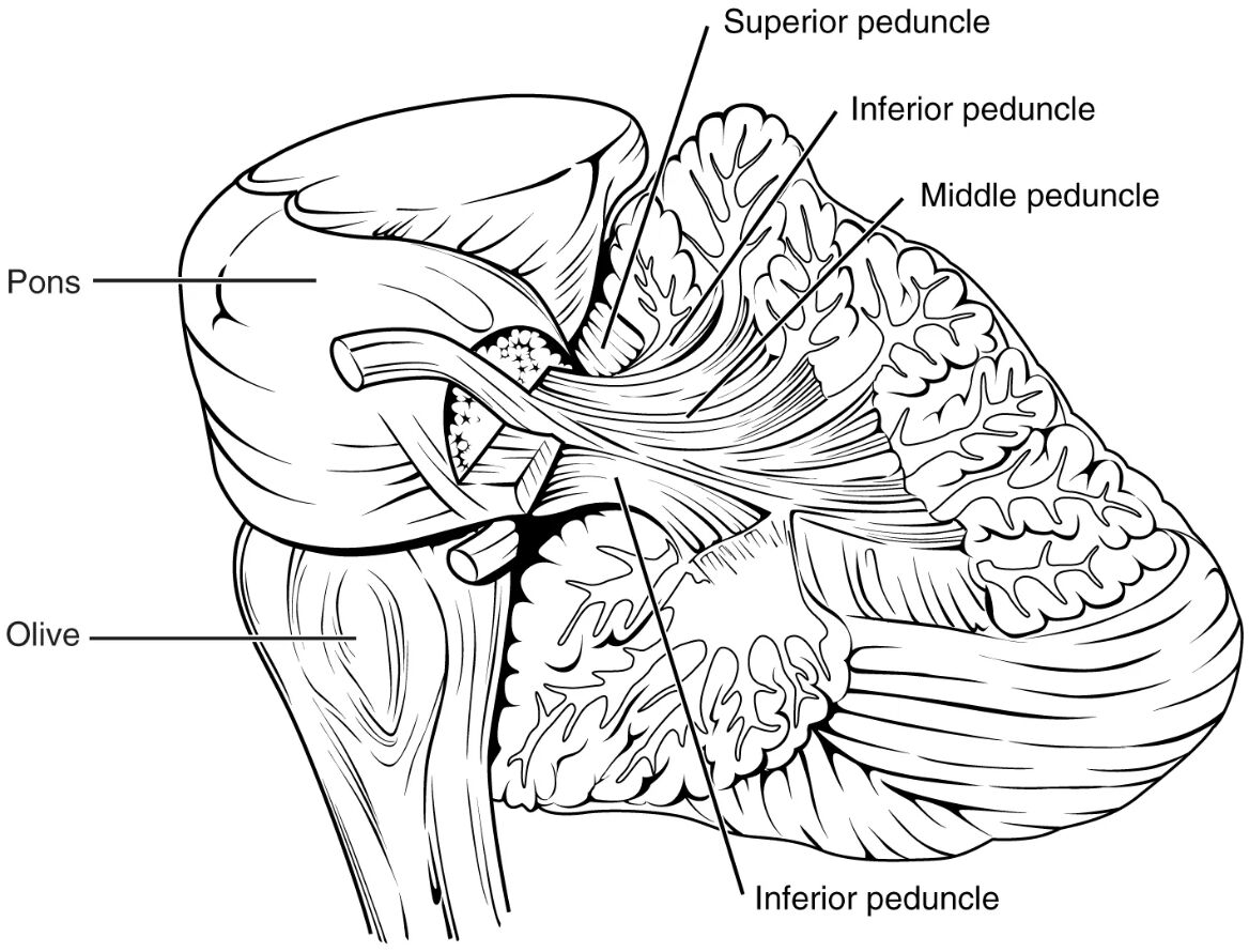
Inferior cerebellar peduncle (ICP) The inferior cerebellar peduncle, arising from the medulla, connects the cerebellum to the spinal cord and lower brainstem. It carries sensory input from the inferior olive, a noticeable bulge on the medulla’s ventral surface, which is crucial for coordinating movement.
Middle cerebellar peduncle (MCP) The middle cerebellar peduncle, located on the ventral surface of the pons, serves as the largest pathway linking the cerebellum to the pons. It transmits corticopontocerebellar fibers, facilitating the relay of information from the cerebral cortex for motor planning.
Superior cerebellar peduncle (SCP) The superior cerebellar peduncle, projecting into the midbrain, is the primary output route for cerebellar efferent fibers to higher brain centers. It carries signals related to motor coordination and balance, crossing over in the decussation of the superior cerebellar peduncles.
Anatomy of the Cerebellar Peduncles
The cerebellar peduncles form critical bridges between the cerebellum and brainstem. This diagram highlights their structural organization.
- The inferior cerebellar peduncle (ICP) integrates sensory data, including proprioception, into cerebellar function.
- The middle cerebellar peduncle (MCP) dominates with its extensive fiber connections from the pons.
- The superior cerebellar peduncle (SCP) serves as the main efferent pathway, linking to the thalamus and cortex.
- These peduncles are located on each side of the cerebellum, forming a symmetrical network.
- White matter tracts within them ensure efficient signal transmission.
Role of the Inferior Cerebellar Peduncle
The ICP plays a key role in sensory integration for the cerebellum. Its connections enhance motor control.
- This peduncle receives climbing fibers from the inferior olive, aiding fine motor adjustments.
- It also carries spinocerebellar tracts, relaying unconscious proprioceptive input.
- The inferior olive’s rhythmic activity supports timing in coordinated movements.
- Damage here can lead to ataxia or tremor due to disrupted sensory feedback.
- Its medullary origin makes it vulnerable to lower brainstem lesions.
Function of the Middle Cerebellar Peduncle
The MCP facilitates communication from the cerebral cortex to the cerebellum. Its extensive fibers support complex tasks.
- This peduncle relays information via pontine nuclei, which process cortical input.
- It enables the cerebellum to refine movements based on planned actions.
- The MCP’s size reflects the volume of corticopontine projections.
- Injury can impair motor learning, affecting skills like writing or playing instruments.
- Its pontine location ties it to higher cognitive-motor integration.
Significance of the Superior Cerebellar Peduncle
The SCP is essential for outputting cerebellar signals to the brain. Its upward projection influences motor execution.
- This peduncle carries dentate nucleus output, crossing in the midbrain for contralateral control.
- It connects to the red nucleus and thalamus, modulating motor pathways.
- The decussation ensures balanced motor responses across the body.
- Lesions here may cause intention tremor or dysmetria due to efferent disruption.
- Its midbrain link supports coordination with visual and auditory systems.
Neural Pathways and Signal Transmission
The peduncles support bidirectional communication within the nervous system. Their design optimizes cerebellar function.
- Afferent fibers in the ICP and MCP bring sensory and cortical data to the cerebellum.
- Efferent fibers in the SCP exit to adjust motor commands in the cortex.
- Myelinated axons within these tracts ensure rapid signal conduction.
- The cerebellum uses this input-output loop for error correction in movement.
- Disruption affects gait, balance, and fine motor skills.
Clinical Relevance of Cerebellar Peduncles
Understanding peduncle anatomy aids in diagnosing neurological conditions. This diagram supports clinical insights.
- ICP damage may result from medullary infarction, leading to sensory ataxia.
- MCP lesions, often from pontine stroke, impair motor planning and execution.
- SCP injuries can cause cerebellar mutism or motor incoordination post-surgery.
- MRI imaging visualizes peduncle integrity, guiding treatment plans.
- Rehabilitation focuses on compensatory strategies for affected functions.
Neurotransmitters and Hormonal Influences
Chemical signaling enhances peduncle function within the cerebellum. These substances modulate neural activity.
- Glutamate excites neurons in the MCP, supporting corticopontine relay.
- GABA inhibits overactive signals in the SCP, refining motor output.
- Dopamine influences motor learning via cerebellar circuits.
- Thyroid hormones like T3 and T4 boost metabolic support for neural transmission.
- Imbalances can exacerbate cerebellar symptoms, necessitating evaluation.
Developmental and Anatomical Variations
Peduncles develop early, with variations influencing function. This diagram reflects a typical adult structure.
- The ICP forms during embryogenesis, linking the medulla by the third trimester.
- The MCP expands with cortical development, peaking in early childhood.
- The SCP matures with cerebellar growth, stabilizing motor control.
- Asymmetry in peduncle size may occur, affecting coordination slightly.
- Aging reduces myelin integrity, potentially slowing peduncle signals.
Advances in Cerebellar Research
Ongoing studies enhance our knowledge of these peduncles. This diagram inspires cutting-edge investigations.
- Diffusion tensor imaging maps white matter tracts within peduncles.
- Deep brain stimulation targets SCP pathways for tremor control.
- Stem cell research explores regenerating damaged ICP fibers.
- Functional MRI assesses MCP activity during motor tasks.
- These innovations promise improved diagnostics and therapies.
In conclusion, this diagram of cerebellar peduncles provides a detailed look at the cerebellum’s vital connections to the brainstem, highlighting the inferior, middle, and superior peduncles’ roles in sensory integration and motor coordination. These pathways form a complex network that ensures smooth movement and balance, offering a window into neurological function. By exploring their anatomy and clinical significance, we can better appreciate their importance and support advancements in treating cerebellar-related disorders, enhancing overall neurological care.

