The anterior view of the right hip joint, with the capsule in place, offers a detailed perspective on a key ball-and-socket joint that supports weight and enables fluid movement. This illustration highlights the ligaments and bony landmarks that work together to maintain stability, especially when standing, providing a clear view of their anatomical significance. Delving into this image enhances your understanding of how the hip joint functions as a cornerstone of lower body mechanics.
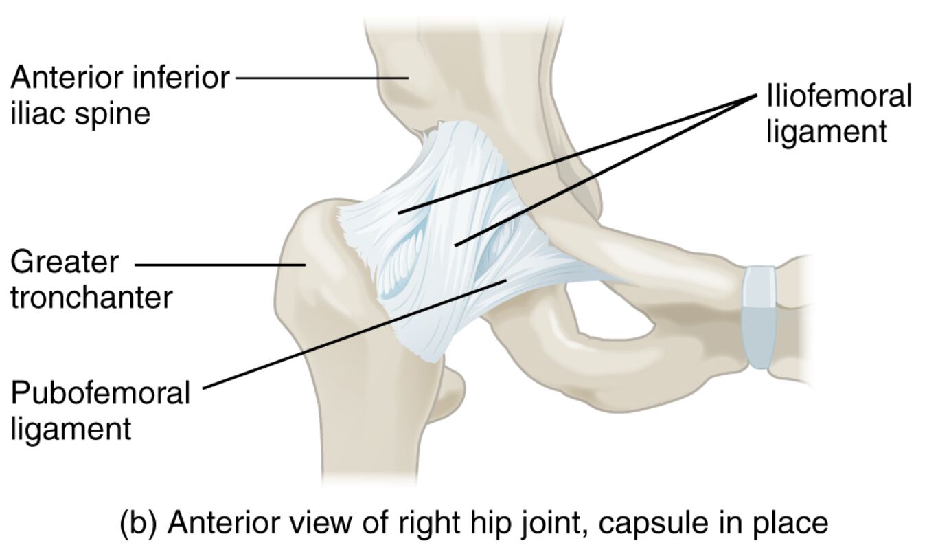
Labeled Parts Explanation
- Anterior inferior iliac spine: This bony projection on the pelvis serves as an attachment point for the rectus femoris muscle, aiding in hip flexion. It also contributes to the structural integrity of the pelvic region.
- Iliofemoral ligament: This strong ligament connects the ilium to the femur, preventing hyperextension of the hip joint. It tightens when standing, pulling the femoral head securely into the acetabulum.
- Greater trochanter: This prominent bony protrusion on the femur provides attachment for hip abductor muscles like the gluteus medius. It plays a crucial role in stabilizing the pelvis during walking.
- Pubofemoral ligament: This ligament extends from the pubis to the femur, limiting excessive abduction and extension of the hip. It works in concert with other ligaments to maintain joint stability.
Introduction to Hip Joint Anatomy
The hip joint is a vital structure in the lower body, functioning as a ball-and-socket joint that supports the body’s weight and allows a wide range of motions. This anterior view illustration of the right hip joint, with the capsule intact, showcases the iliofemoral and pubofemoral ligaments alongside key bony landmarks like the greater trochanter. A thorough exploration of these components reveals how they collaborate to ensure stability, particularly when upright.
- Provides a clear overview of the hip’s anatomical layout.
- Highlights the joint’s importance in weight-bearing activities.
Role of Ligaments in Hip Stability
Ligaments are essential for securing the hip joint, with each serving a distinct function. The iliofemoral ligament, known for its strength, prevents the hip from overextending, becoming taut when standing to anchor the femoral head. The pubofemoral ligament complements this by restricting abduction, ensuring the joint remains aligned during movement.
- Explains how ligament tension supports the hip during standing.
- Details the coordinated action of ligaments in joint protection.
Bony Landmarks and Muscle Attachments
The hip’s skeletal structure includes significant features that support movement and stability. The greater trochanter acts as a anchor for muscles that stabilize the pelvis, crucial for maintaining balance while walking. The anterior inferior iliac spine provides a point for muscle attachment, enhancing hip flexion and overall joint function.
- Describes the greater trochanter’s role in pelvic stability.
- Emphasizes the anterior inferior iliac spine’s contribution to muscle action.
Clinical Relevance and Functional Insights
Understanding the hip’s anterior anatomy is key to recognizing potential issues, such as ligament strains or joint instability. The iliofemoral ligament’s strength is critical in preventing dislocations, while the pubofemoral ligament helps avoid excessive lateral motion. This knowledge can inform rehabilitation strategies to restore hip function after injury.
- Offers insight into common hip stability challenges.
- Suggests the value of anatomical understanding in treatment planning.
Conclusion
The anterior view of the right hip joint illustrates a remarkable synergy of ligaments and bony structures that ensure its stability and functionality. From the robust iliofemoral ligament to the supportive greater trochanter, each element plays a vital role in supporting the body’s daily movements. Exploring this anatomy not only deepens your appreciation of the hip’s complexity but also equips you with the insight to maintain its health effectively.

