Medium-sized veins are key players in the circulatory system, facilitating the return of deoxygenated blood to the heart with a structure that balances flexibility and support. This image presents a sectional view of a medium-sized vein, highlighting its layered anatomy and the presence of valves that ensure efficient blood flow, offering a clear insight into its functional design.
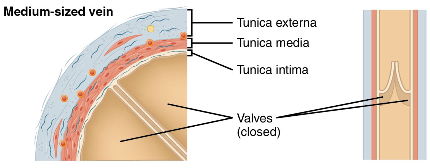
Tunica externa The tunica externa is the outermost layer, composed of dense connective tissue that provides structural support and anchors the vein to surrounding tissues. It contains small blood vessels and nerves, ensuring the vein remains nourished and responsive to physiological demands.
Tunica media The tunica media is the middle layer, featuring a thin layer of smooth muscle cells and elastic fibers that allow the vein to stretch and contract. This layer contributes to venous tone, aiding in the regulation of blood flow and volume.
Tunica intima The tunica intima is the innermost layer, lined with endothelial cells that minimize friction and support smooth blood flow. In medium-sized veins, this layer includes valves that prevent backflow, a critical feature for venous return.
Valve The valve is a flap-like structure within the tunica intima that opens and closes to ensure unidirectional blood flow toward the heart. When closed, it prevents backflow, particularly against gravity in the lower extremities.
Smooth muscle cell in tunica media Smooth muscle cells in the tunica media help regulate the vein’s diameter by contracting or relaxing. These cells assist in maintaining venous pressure and supporting blood return during physical activity.
The Role of Medium-Sized Veins in Circulation
Medium-sized veins serve as an essential link between smaller venules and larger veins in the circulatory system. Their structure supports the body’s ability to manage blood flow effectively.
- The tunica externa provides a protective and flexible framework for the vein.
- The tunica media allows the vein to adapt to changes in blood volume and pressure.
- Valves ensure blood moves efficiently toward the heart, reducing the risk of pooling.
- This design facilitates the transport of deoxygenated blood, including hormones like T3 and T4 from the thyroid gland.
Anatomical Layers of Medium-Sized Veins
The sectional view reveals the distinct layers that compose a medium-sized vein. Each layer plays a specific role in maintaining the vein’s integrity and function.
- The tunica intima forms a smooth inner lining, with valves to prevent reverse flow.
- The tunica media contains smooth muscle cells that regulate vessel diameter.
- The tunica externa offers structural support and houses nourishing vessels.
- Together, these layers balance flexibility and strength for venous health.
Physiological Functions and Importance
The anatomy of medium-sized veins enables them to perform vital physiological roles. Their design ensures efficient blood return and adaptation to bodily needs.
- The valve opens during muscle contraction to propel blood upward and closes to prevent backflow.
- Smooth muscle cells in tunica media contract to aid venous return, especially in the legs.
- The tunica intima supports the exchange of gases and nutrients as blood moves.
- This adaptability is crucial for maintaining circulation during rest or activity.
Clinical Significance of Medium-Sized Vein Anatomy
Understanding the structure of medium-sized veins provides insights into potential health issues. Changes in these layers can indicate or contribute to circulatory problems.
- Weak valves can lead to varicose veins, causing discomfort and swelling.
- Thickening of the tunica media may occur in chronic venous insufficiency.
- Damage to the tunica intima increases the risk of deep vein thrombosis.
- Research into vein anatomy supports treatments for vascular diseases.
Comparison with Other Blood Vessels
Medium-sized veins differ from arteries, large veins, and capillaries due to their unique structure. This comparison highlights their specific role in the circulatory system.
- Unlike arteries, the tunica media in veins is thinner, reflecting lower pressure needs.
- Large veins lack the valves found in medium-sized veins, relying on size for flow.
- Capillaries lack the layered structure, focusing on exchange rather than transport.
- The presence of smooth muscle cells in tunica media distinguishes medium-sized veins from venules.
Maintenance and Regulation of Medium-Sized Veins
The body employs mechanisms to maintain the health and function of medium-sized veins. These processes ensure optimal performance under varying conditions.
- The tunica externa is nourished by small vessels, supporting its connective tissue.
- Smooth muscle cells in tunica media respond to neural signals to adjust tone.
- The valve opens and closes dynamically with blood pressure changes.
- Hormonal influences help regulate the tunica intima to prevent clotting.
In conclusion, the sectional view of a medium-sized vein, as depicted in this image, showcases a well-designed structure for efficient blood return. With its tunica externa, tunica media, tunica intima, valve, and smooth muscle cells in tunica media, this vein type exemplifies the circulatory system’s ability to adapt to diverse physiological demands. Exploring these features deepens our understanding of venous anatomy and its critical role in sustaining health.

