Uncover the intricate dance of the heart valves during the critical phase of ventricular contraction, as vividly illustrated in this detailed image. This exploration reveals how these vital structures meticulously regulate blood flow, ensuring its unidirectional movement and efficient circulation throughout the body. Understanding the synchronized opening and closing of heart valves is fundamental to grasping cardiac function and recognizing the signs of potential valvular disease.
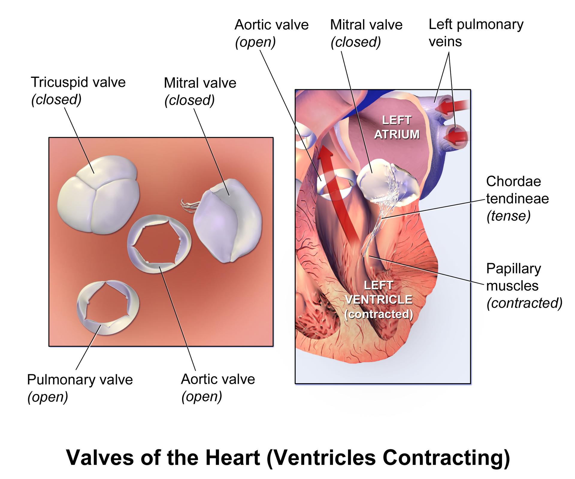
Tricuspid valve (closed): The tricuspid valve is positioned between the right atrium and the right ventricle. During ventricular contraction, it is closed to prevent the backflow of deoxygenated blood into the right atrium, ensuring efficient forward propulsion into the pulmonary artery.
Mitral valve (closed): The mitral valve, also known as the bicuspid valve, is located between the left atrium and the left ventricle. When the ventricles contract, this valve is closed to prevent the backflow of oxygenated blood into the left atrium, ensuring that blood is directed into the aorta.
Pulmonary valve (open): The pulmonary valve is situated at the exit of the right ventricle, leading into the pulmonary artery. During ventricular contraction, this valve is open, allowing deoxygenated blood to be forcefully ejected from the right ventricle into the pulmonary circulation.
Aortic valve (open): The aortic valve is located at the exit of the left ventricle, leading into the aorta. As the ventricles contract, this valve is open, permitting the forceful ejection of oxygenated blood from the left ventricle into the systemic circulation.
Left pulmonary veins: The left pulmonary veins are responsible for carrying oxygenated blood from the left lung back to the left atrium of the heart. These veins are crucial for completing the pulmonary circuit and delivering enriched blood for systemic distribution.
Left Atrium: The left atrium is the upper left chamber of the heart that receives oxygenated blood from the lungs via the pulmonary veins. During ventricular contraction, it is relaxed and filling with blood, preparing for the next cardiac cycle.
Chordae tendineae (tense): The chordae tendineae are tough, fibrous cords that connect the cusps of the atrioventricular (mitral and tricuspid) valves to the papillary muscles. When the ventricles contract, they become tense, preventing the valve cusps from prolapsing back into the atria.
Papillary muscles (contracted): Papillary muscles are located in the ventricles and are responsible for pulling on the chordae tendineae during ventricular contraction. Their contraction helps to keep the atrioventricular valves closed, preventing backflow of blood into the atria.
Left Ventricle (contracted): The left ventricle is the strongest chamber of the heart, responsible for pumping oxygenated blood into the aorta and throughout the body. During its contracted state, it generates significant pressure to propel blood into systemic circulation.
The human heart operates through a precisely synchronized series of contractions and relaxations, known as the cardiac cycle. A critical phase within this cycle is ventricular contraction, or systole, where the ventricles forcefully expel blood into the pulmonary artery and aorta. This image provides an exceptional view of how the heart valves function during this powerful pumping action, demonstrating the coordinated opening and closing that directs blood flow efficiently. The atrioventricular valves (tricuspid and mitral) are shown closed to prevent backflow into the atria, while the semilunar valves (pulmonary and aortic) are open to allow blood ejection.
The mechanical action of the heart valves is crucial for maintaining a unidirectional flow of blood, preventing regurgitation and ensuring optimal cardiac output. During ventricular contraction, the significant pressure generated would force blood back into the atria if the mitral and tricuspid valves did not close tightly. This closure is actively supported by the chordae tendineae and papillary muscles, which become tense and contract respectively, holding the valve leaflets in place. Simultaneously, the rising pressure within the ventricles forces open the aortic and pulmonary valves, allowing blood to exit the heart.
Malfunctions of these valves can lead to serious cardiovascular conditions. For instance, if the mitral valve fails to close completely during ventricular contraction, a condition known as mitral regurgitation occurs. This allows blood to leak back into the left atrium, increasing the workload on the heart and potentially leading to symptoms like shortness of breath and fatigue. Similarly, aortic stenosis, a narrowing of the aortic valve, impedes the flow of blood from the left ventricle into the aorta, requiring the heart to work harder to pump blood.
- Heart sounds, “lub-dub,” are primarily caused by the closing of the heart valves.
- The mitral and tricuspid valves close first (“lub”), followed by the aortic and pulmonary valves (“dub”).
- Each valve has a unique structure tailored to its function and location.
- Valve replacement surgery is a common treatment for severe valvular diseases.
This intricate interplay of contracting muscles, tensing cords, and opening and closing valves underscores the remarkable efficiency of the human heart. The clarity of this illustration provides an invaluable educational tool for understanding the fundamental principles of cardiovascular physiology. A healthy functioning heart, with its valves working in perfect harmony, is paramount for sustaining life and overall well-being.
The detailed illustration of heart valve dynamics during ventricular contraction offers a profound insight into the precision and efficiency of the cardiovascular system. Each valve’s specific action, coordinated by surrounding structures like the chordae tendineae and papillary muscles, ensures the unidirectional flow of blood. This understanding is critical for medical diagnosis, treatment of valvular heart diseases, and for anyone seeking to comprehend the fundamental mechanics of the human heart.

