The human skin is a complex, multi-layered organ that serves as the primary interface between the internal physiological environment and the external world. At the very edge of this interface lies the stratum corneum, a thin but incredibly resilient layer that defines the skin’s protective capabilities. In modern dermatology and pharmacological research, analyzing this layer has become a cornerstone for understanding disease progression and drug efficacy. Non-invasive sampling techniques allow clinicians to peer into the molecular health of a patient without the need for traditional, invasive biopsies. By harvesting the superficial cells of the epidermis, researchers can quantify inflammatory markers, lipid profiles, and protein concentrations that provide a comprehensive snapshot of the body’s largest organ.
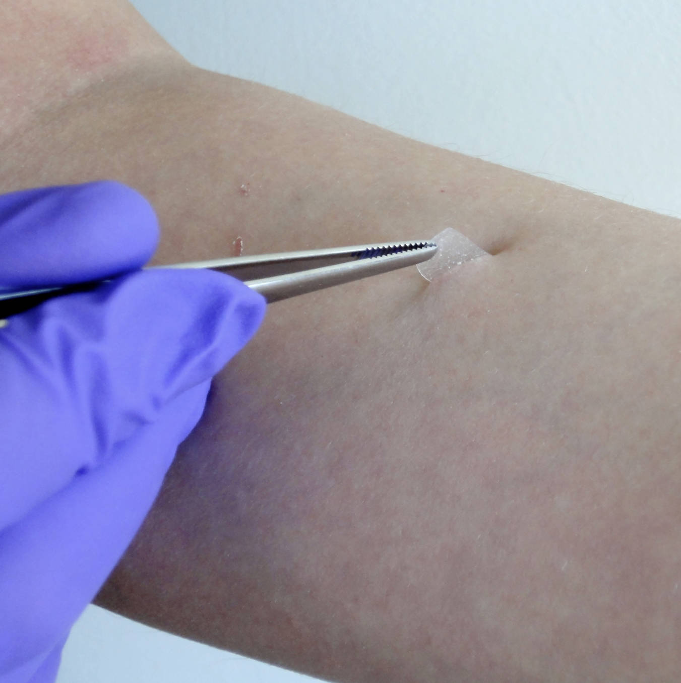
Forceps: In this procedure, precision forceps are utilized to grasp the edge of the sampling material with minimal physical interference. This tool ensures that the removal process is steady and controlled, preventing accidental tearing of the delicate skin sample.
Adhesive Strip: A specialized medical-grade tape is applied to the surface of the skin to facilitate the collection of biological material. When peeled back, this strip lifts a thin layer of cells, which can then be processed for laboratory analysis using various biochemical assays.
Human Skin: The forearm, shown here, is a frequent site for cutaneous research due to its low hair density and relatively stable vascularity. The skin surface must be properly prepared to ensure that the collected sample accurately represents the underlying physiological state.
Gloved Hand: The presence of a sterile nitrile glove highlights the importance of maintaining an aseptic environment during the sampling process. This protocol prevents the transfer of external contaminants or the practitioner’s own oils and microbes to the critical diagnostic sample.
The Anatomy and Physiology of the Stratum Corneum
To appreciate the value of sampling this layer, one must understand its unique architecture. Often described by the ‘bricks and mortar’ model, the stratum corneum consists of flattened, dead cells known as corneocytes embedded in a lipid-rich extracellular matrix. While these cells have lost their nuclei and metabolic activity, they remain structurally vital, providing the mechanical strength and chemical resistance necessary to thwart pathogens and prevent excessive moisture loss. The ‘mortar’ consists of ceramides, cholesterol, and free fatty acids, which together form a waterproof seal that is essential for life in a terrestrial environment.
The thickness of this layer varies significantly across different anatomical regions of the body, ranging from the extremely thin skin of the eyelids to the thickened, protective pads of the palms and soles. Despite its apparent simplicity, the stratum corneum is a biologically active site for desquamation, where cells are shed and replaced in a highly regulated cycle. Dysregulation of this cycle or changes in the chemical composition of the lipids can lead to a breakdown of the epidermal barrier, resulting in clinical conditions such as atopic dermatitis or xerosis.
The Methodology of Tape Stripping in Research
The image provided demonstrates a classic example of tape stripping, a widely used method for sequentially removing layers of the stratum corneum. This technique is valued for its non-invasive nature and its ability to provide a depth-resolved analysis of the skin. By applying multiple strips to the same site, researchers can determine how the concentration of a topically applied drug changes as it penetrates deeper into the epidermis. Each strip captures approximately one micrometer of tissue, allowing for a precise mapping of the skin’s absorption characteristics.
Standardization is key to obtaining reliable data from this procedure. Factors such as the amount of pressure applied to the tape, the duration of application, and the speed at which the tape is removed can all influence the amount of protein harvested. Advanced clinical settings often use automated devices to ensure that pressure is consistent across all subjects. Once the strips are collected, they undergo extraction processes to release the biological material, which is then analyzed using techniques like liquid chromatography-mass spectrometry or enzyme-linked immunosorbent assays (ELISA).
Diagnostic Utility and Biomarker Discovery
One of the most exciting frontiers in cutaneous medicine is the use of stratum corneum sampling for biomarker discovery. Historically, monitoring internal inflammation required blood draws or tissue biopsies, but we now know that many systemic signals are reflected in the skin’s outermost layer. For instance, cytokines such as interleukin-1 and interleukin-8 can be measured from a single tape strip, providing an early warning of an impending inflammatory flare in patients with chronic skin disorders. This ‘molecular biopsy’ is particularly beneficial for pediatric patients, for whom invasive procedures are often traumatic and difficult to manage.
Beyond inflammation, sampling allows for the study of the skin’s natural moisturizing factors (NMFs). These small, water-holding molecules are the primary regulators of skin hydration. By quantifying NMF levels through sampling, clinicians can tailor topical treatments to the specific needs of a patient’s skin. This personalized approach to dermatology ensures that interventions are both effective and efficient, reducing the trial-and-error often associated with treating dry or sensitive skin conditions.
Assessing Barrier Function and Transepidermal Water Loss
A primary goal of evaluating the stratum corneum is to measure the integrity of the skin barrier. A weakened barrier is often characterized by a spike in transepidermal water loss (TEWL), a metric that quantifies the amount of water evaporating through the skin surface. High TEWL values are a hallmark of impaired skin and serve as a sensitive indicator of damage, even before visible symptoms like redness or scaling appear. Sampling the stratum corneum in conjunction with TEWL measurements provides a powerful dual-diagnostic approach to assess both the structural and functional status of the skin.
In occupational health, these techniques are used to screen workers exposed to harsh chemicals or irritants. By monitoring the molecular changes in the skin before clinical dermatitis develops, employers and medical teams can implement preventive measures to protect at-risk individuals. This proactive stance on skin health reduces the incidence of long-term disability and improves the overall well-being of the workforce, highlighting the broad public health implications of these simple sampling techniques.
Future Directions in Skin Sampling Technology
The evolution of skin sampling is moving toward even more refined and user-friendly technologies. Micro-needle patches and hydrogel-based samplers are currently in development, promising to capture a wider array of biomarkers with even greater sensitivity. These innovations aim to make skin testing as common and accessible as blood glucose monitoring, potentially allowing patients to track their own skin health at home using smartphone-integrated diagnostic tools. The integration of artificial intelligence with molecular data from skin samples could soon lead to predictive models that anticipate disease outbreaks weeks in advance.
Furthermore, the pharmaceutical industry is increasingly relying on these methods to accelerate drug development. By providing immediate feedback on how a new formula interacts with the stratum corneum, researchers can refine their designs in the early stages of clinical trials. This reduces the time and cost associated with bringing new dermatological therapies to market. As our understanding of the skin’s molecular landscape continues to grow, the humble tape strip remains a vital bridge between the laboratory and the bedside, transforming the way we perceive, treat, and maintain the health of our skin.

