The human lower extremity is an architectural masterpiece of biological engineering, designed to facilitate upright locomotion while simultaneously managing the massive gravitational loads of the upper body. From the robust structure of the pelvic girdle down to the intricate arrangement of the foot, every segment of the leg is optimized for stability, power, and precision. This system is not merely a collection of bones and muscles; it is a dynamic network of vascular channels, nerve pathways, and connective tissues that work in a synchronized rhythm to allow us to move through the world. Understanding the external landmarks and underlying anatomy of the leg is fundamental to recognizing how our bodies adapt to physical stress and where potential vulnerabilities may lie. Whether in a clinical setting or an athletic environment, appreciating the complexities of the lower limb provides the foundation for effective diagnostic assessment and long-term musculoskeletal health.
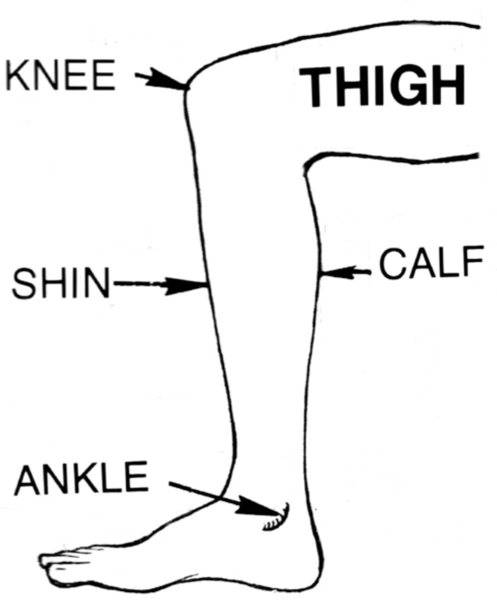
KNEE: The knee represents the largest and most complex joint in the human body, acting as a pivotal hinge between the thigh and the lower leg. It involves the articulation of the femur, tibia, and patella, supported by a sophisticated network of ligaments and menisci to manage high weight-bearing loads.
THIGH: The thigh is the proximal region of the lower limb, extending from the hip to the knee and housing the femur, the body’s longest and strongest bone. It contains major muscle groups such as the quadriceps and hamstrings, which are essential for powerful movements like running and jumping.
SHIN: The shin refers to the anterior portion of the lower leg, specifically characterized by the subcutaneous border of the tibia. This area is minimally protected by muscle or fat, making the underlying bone particularly susceptible to direct trauma and common inflammatory conditions like medial tibial stress syndrome.
CALF: The calf is the fleshy posterior region of the lower leg, primarily composed of the gastrocnemius and soleus muscles. These muscles converge into the Achilles tendon and are fundamental for plantarflexion of the foot, providing the necessary power for the propulsion phase of the gait cycle.
ANKLE: The ankle is the complex synovial joint that connects the foot to the leg, formed by the distal ends of the tibia and fibula meeting the talus bone. It allows for critical movements like dorsiflexion and eversion, providing the stability and flexibility required to navigate uneven terrain.
Structural Anatomy of the Thigh and Knee Complex
The thigh is defined by the presence of the femur, a bone so structurally sound that it can withstand forces several times an individual’s body weight. Surrounding this bone are three main compartments of muscles: the anterior, medial, and posterior. The anterior compartment is dominated by the quadriceps femoris, which serves as the primary extensor of the knee. On the medial side, the adductor group facilitates the inward movement of the leg, while the posterior compartment contains the hamstrings, which are vital for knee flexion and hip extension. This balanced muscular arrangement is what allows for the explosive power seen in sprinters and the steady endurance of long-distance walkers.
Moving distally, the knee joint acts as the interface between the thigh and the lower leg. It is a modified hinge joint, meaning it primarily performs flexion and extension but also allows for a small degree of rotation when flexed. The internal integrity of the knee is maintained by the cruciate ligaments (ACL and PCL), which prevent the tibia from sliding too far forward or backward relative to the femur. Protecting the bony surfaces is the **articular cartilage**, a smooth, slippery tissue that reduces friction and absorbs shock. When this cartilage wears down, it often leads to the development of osteoarthritis, a common condition that significantly impacts mobility in aging populations.
The Lower Leg: Biomechanics of the Shin and Calf
The lower leg, often called the crural region, is composed of the tibia (shinbone) and the fibula. While the tibia is the main weight-bearer of the **skeletal system** in this region, the fibula acts as a lateral stabilizer and an attachment point for various muscles. The shin is particularly notable in medical diagnostics because the anterior border of the tibia is subcutaneous, meaning there is very little tissue between the bone and the skin. This makes the shin a frequent site of bruises and lacerations, but also allows clinicians to easily palpate the bone for fractures or irregularities.
In contrast, the posterior part of the lower leg, the calf, is a dense powerhouse of muscle. The gastrocnemius and soleus muscles are the primary drivers of plantarflexion, the movement of pointing the toes downward. This action is the primary force behind the “push-off” during walking or running. These muscles merge to form the Achilles tendon, which inserts into the heel bone (calcaneus). Because of the high tension placed on this tendon during athletic activities, it is a frequent site of inflammatory injuries or even complete ruptures. The relationship between the anterior shin muscles and the posterior calf muscles must be carefully balanced to prevent foot drop or gait abnormalities.
The Ankle: Nexus of Stability and Proprioception
The ankle joint, or talocrural joint, is where the leg meets the foot. It is formed by the medial malleolus of the tibia, the lateral malleolus of the fibula, and the body of the talus bone. This mortise-and-tenon structure is designed for incredible stability. Surrounding the joint are several key ligaments, most notably the deltoid ligament on the medial side and the anterior talofibular ligament (ATFL) on the lateral side. Lateral ankle sprains, involving the ATFL, are the most common musculoskeletal injury in both athletes and the general population, usually occurring during a sudden inversion of the foot.
Beyond its mechanical function, the ankle is a vital center for **proprioception**, the body’s internal sense of its position in space. Sensory receptors in the ligaments and tendons of the ankle send constant feedback to the brain, allowing for instantaneous adjustments in balance when walking on uneven ground. This neurological feedback loop is what allows humans to maintain a bipedal stance with minimal conscious effort. When this system is compromised by injury or neurological disease, the risk of falls and subsequent fractures increases significantly, particularly in elderly patients whose reflex speeds may be diminished.
Common Pathologies and Clinical Considerations
Injuries to the lower extremity are diverse, ranging from acute trauma to chronic overuse syndromes. In the knee, the anterior cruciate ligament (ACL) is particularly vulnerable to tearing during pivoting movements. In the shin, athletes often struggle with “shin splints,” or medial tibial stress syndrome, which results from the inflammation of the muscles and tendons along the tibia. This is often caused by repetitive stress on hard surfaces or improper footwear. Stress fractures of the tibia are a more severe manifestation of this same repetitive loading, requiring significant periods of rest for the bone to remodel and heal.
The **biomechanics** of the lower limb can also be affected by systemic conditions. Peripheral artery disease (PAD) can cause claudication, or cramping pain in the calves during exercise, due to restricted blood flow. Similarly, deep vein thrombosis (DVT) frequently occurs in the large veins of the calf, posing a serious risk of pulmonary embolism if the clot dislodges. Clinical examination of the lower extremity often includes checking for swelling (edema), assessing peripheral pulses, and testing dermatomes to ensure the neurological pathways from the lumbar spine are intact. A thorough understanding of these anatomical regions allows for a more targeted approach to both surgical and conservative treatments.
Preventative Care and Long-term Musculoskeletal Health
Maintaining the health of the lower extremity is a lifelong process that centers on balance, strength, and proper mechanics. Regular weight-bearing exercise is essential for maintaining bone density and preventing osteoporosis. Strengthening the muscles around the knee and ankle provides a protective sleeve that reduces the stress on the joints themselves. Flexibility is equally important; tight calf muscles can lead to plantar fasciitis or Achilles tendonitis by altering the natural mechanics of the foot during movement.
Nutritional support, including adequate calcium and Vitamin D, ensures that the bones of the thigh, shin, and ankle remain strong. Additionally, wearing supportive, well-fitted footwear can prevent many of the mechanical issues that lead to chronic pain in the shin and knee. As we age, maintaining the integrity of the lower limb is synonymous with maintaining independence. By understanding the anatomy from the thigh down to the ankle, we can better appreciate the incredible demands we place on our legs every day and take the necessary steps to preserve their function for decades to come.

