The heart’s intricate internal structure is essential for its role as a pump, and this image showcases key components that ensure proper valve function. This frontal section highlights the chordae tendineae and papillary muscles, which are critical for supporting the tricuspid and mitral valves, offering a clear view of their anatomical arrangement. Delving into this image provides a deeper appreciation of how these structures maintain efficient blood flow within the heart.
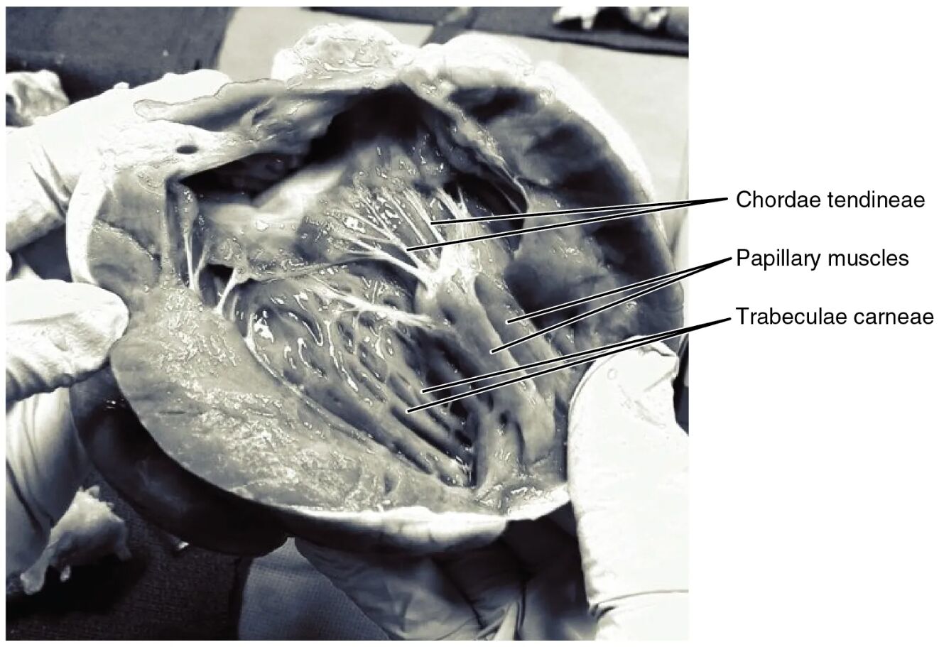
Chordae tendineae: The chordae tendineae are tough, fibrous cords that connect the papillary muscles to the tricuspid and mitral valves, preventing valve prolapse during heart contraction. These cords ensure the valves close properly, maintaining unidirectional blood flow from the atria to the ventricles.
Papillary muscles: The papillary muscles are conical projections of the ventricular wall that anchor the chordae tendineae, providing structural support to the valves. They contract during systole to tension the chordae, stabilizing the valves against the pressure of blood flow.
Trabeculae carneae: The trabeculae carneae are irregular muscular ridges lining the inner surface of the ventricles, enhancing the heart’s contractile strength. They also serve as attachment points for the papillary muscles, contributing to the overall structural integrity of the heart.
Anatomical Structure of the Heart’s Internal Components
The heart’s internal anatomy includes specialized structures that support its pumping mechanism. This image provides a detailed look at the interplay between these critical elements.
- The chordae tendineae form a network that anchors the valves, ensuring they withstand the force of ventricular contraction.
- Papillary muscles arise from the ventricular myocardium, playing a dynamic role in valve function during each heartbeat.
- Trabeculae carneae create a textured interior, increasing the surface area for muscle contraction and oxygen delivery.
- Together, these structures work seamlessly to prevent backflow and maintain cardiac efficiency.
Their arrangement reflects the heart’s evolutionary adaptation to high-pressure environments.
Functional Roles in Heart Performance
These anatomical features are integral to the heart’s ability to pump blood effectively. The image illustrates their active roles during the cardiac cycle.
- Chordae tendineae tighten during systole, pulling the valve leaflets closed to prevent regurgitation.
- Papillary muscles contract synchronously with the ventricles, providing the necessary tension to support the valves.
- Trabeculae carneae enhance the ventricle’s contractile force, aiding in efficient blood ejection.
- This coordination ensures optimal blood flow from the right atrium to the pulmonary circuit and the left atrium to the systemic circulation.
Any disruption in this system can lead to significant cardiac issues.
Physical Characteristics and Clinical Relevance
The physical properties of these structures contribute to their functionality within the heart. Their appearance and resilience are key to understanding potential complications.
- Chordae tendineae are thin yet strong, capable of withstanding repeated stress without tearing.
- Papillary muscles vary in size and number, adapting to the workload of each ventricle.
- Trabeculae carneae’s irregular pattern increases muscle mass, supporting the heart’s endurance.
- Damage to these structures, such as from mitral valve prolapse, can result in heart murmurs or heart failure.
Regular echocardiograms can assess their condition and guide treatment.
Importance in Cardiac Health
Maintaining the integrity of chordae tendineae, papillary muscles, and trabeculae carneae is vital for long-term cardiac health. Their role extends beyond mechanics to overall circulatory stability.
- Strong chordae tendineae prevent valve leakage, preserving cardiac output.
- Healthy papillary muscles ensure valve competence, reducing the risk of regurgitation.
- Robust trabeculae carneae support ventricular function, especially under increased demand.
- Surgical repair or replacement may be needed if these structures are compromised, such as in rheumatic heart disease.
Monitoring through clinical exams helps detect early signs of dysfunction.
Conclusion
This image of the heart’s internal anatomy highlights the essential roles of the chordae tendineae, papillary muscles, and trabeculae carneae in supporting valve function and maintaining blood flow. Their coordinated action ensures the heart operates efficiently, protecting against conditions that could impair circulation. This detailed view fosters a greater understanding of cardiac anatomy and underscores the importance of preserving these structures for optimal heart health.

