The microscopic study of arteries reveals the intricate cellular architecture that enables them to transport oxygenated blood under high pressure from the heart to the body’s tissues. This image, captured under a microscope, showcases the tunica intima, tunica media, tunica adventitia, and endothelial cells, highlighting the specialized layers that ensure arterial resilience and function.
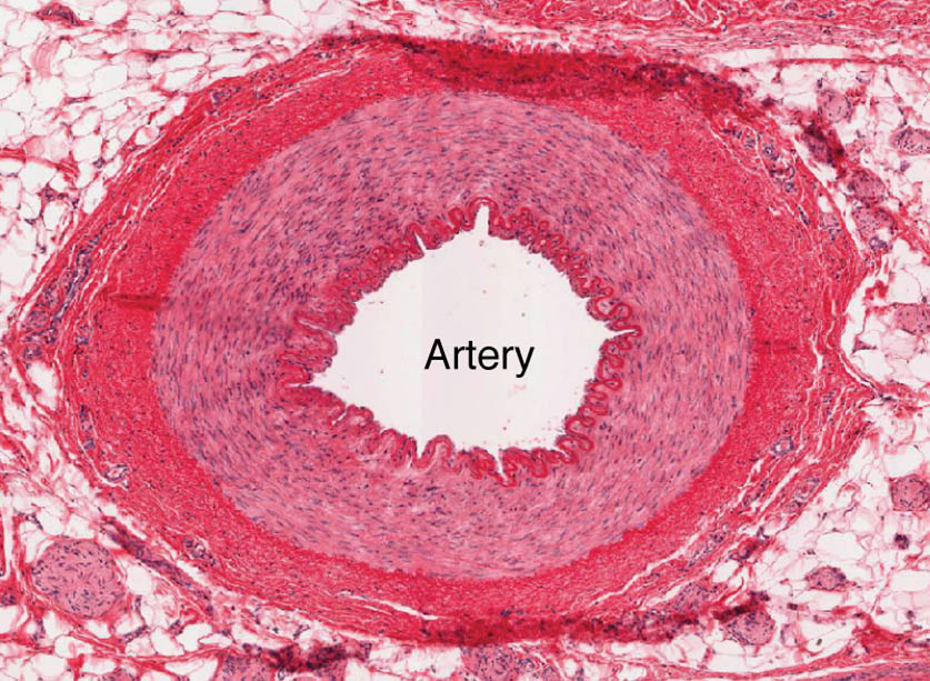
Tunica intima Tunica intima is the innermost layer, featuring a single layer of endothelial cells that line the arterial lumen and reduce friction. This layer includes a subendothelial layer and a prominent internal elastic lamina, which provides elasticity to withstand the pulsatile blood flow.
Tunica media Tunica media forms the thick middle layer, composed of smooth muscle cells and multiple elastic lamellae that regulate vessel diameter and absorb pressure. This robust structure enables the artery to handle systolic pressures up to 120 mmHg while maintaining structural integrity.
Tunica adventitia Tunica adventitia is the outer layer, consisting of connective tissue and collagen fibers that anchor the artery to surrounding tissues. This layer contains vasa vasorum, small vessels that supply nutrients to the arterial wall, particularly in larger arteries.
Endothelial cells Endothelial cells line the tunica intima, appearing as flattened cells under the microscope, and play a key role in regulating blood flow and preventing clotting. These cells release nitric oxide to promote vasodilation and respond to shear stress, ensuring vascular health.
The Role of Microscopic Layers in Arterial Function
This analysis uncovers how each layer supports the artery’s high-pressure role. The microscopic view provides a detailed look at their contributions to circulation.
- Tunica Intima Function: The endothelial cells form a smooth surface, minimizing turbulence and preventing platelet adhesion. The internal elastic lamina stretches with each heartbeat, enhancing flexibility.
- Tunica Media Role: The smooth muscle allows vasoconstriction and vasodilation, controlled by the autonomic nervous system. Elastic lamellae store energy during systole, releasing it to maintain pressure during diastole.
- Tunica Adventitia Support: The collagen network provides tensile strength, preventing arterial rupture. The vasa vasorum ensures oxygen and nutrients reach the outer layers of thick-walled arteries.
- Endothelial Cell Activity: These cells secrete factors like prostacyclin to inhibit clotting, protecting the vessel. They also adjust vessel tone in response to blood flow changes.
Anatomical Details Under the Microscope
The microscopic image reveals the fine structure of arterial layers with high clarity. The magnification highlights the density and organization of cellular components.
This detailed perspective aids in understanding arterial resilience. It serves as a valuable tool for studying vascular anatomy and pathology.
- Tunica Intima Composition: The endothelium is supported by a thin subendothelial layer, with the internal elastic lamina appearing as a dark band. This structure varies in thickness across different artery sizes.
- Tunica Media Thickness: The layer shows multiple concentric elastic lamellae, interspersed with smooth muscle, especially in elastic arteries like the aorta. The density of elastic tissue decreases in smaller muscular arteries.
- Tunica Adventitia Variation: Collagen fibers dominate this layer, appearing as a dense network under the microscope. The vasa vasorum are visible as small, branching vessels within larger arteries.
- Endothelial Cell Morphology: These cells appear elongated due to high pressure, with visible nuclei indicating active function. Their alignment follows the direction of blood flow.
Physiological Functions of Arterial Layers
The physiological roles of these layers are tailored to handle high-pressure blood flow. Their design ensures effective oxygen and nutrient delivery to tissues.
Each layer contributes uniquely to arterial performance. This functionality supports the body’s cardiovascular demands.
- Blood Pressure Regulation: The tunica media’s elastic fibers absorb the systolic peak, smoothing the pressure wave. This protects smaller vessels from damage during each heartbeat.
- Vasomotor Control: Smooth muscle in the tunica media responds to adrenaline, adjusting vessel diameter during exercise. This regulation balances blood flow to active tissues.
- Oxygen Delivery: Arteries carry oxygenated blood rich in glucose and oxygen, facilitated by the tunica intima’s smooth surface. This ensures efficient nutrient supply to capillary beds.
- Pulse Propagation: The elastic recoil of the tunica media and intima propagates the pulse wave, aiding peripheral perfusion. This mechanism maintains systemic pressure.
Comparative Anatomy with Other Vessels
The microscopic view of an artery contrasts with veins and capillaries. These differences reflect the artery’s unique role in high-pressure circulation.
The image emphasizes the artery’s distinct layering. This contrast is key to understanding vascular diversity.
- Artery vs. Vein: Arteries have a thick tunica media with elastic lamellae, while veins feature a thicker tunica adventitia and valves. This reflects their pressure and flow differences.
- Artery vs. Capillary: The tunica media thins as arteries transition to arterioles, reducing to a single endothelial layer in capillaries. This shift maximizes exchange surfaces.
- Elasticity Focus: The tunica media’s elastic fibers are prominent in arteries, aiding recoil. Veins rely on collagen, appearing less elastic under the microscope.
- Staining Techniques: Hematoxylin stains nuclei blue, while eosin highlights elastic fibers in the tunica media. This differentiation aids in identifying arterial tissue.
Clinical and Research Perspectives
Microscopic views of arteries provide valuable insights for medical practice. The layered structure is a focus for studying cardiovascular health and disease.
Advances in microscopy and histology enhance these studies, offering diagnostic tools. These efforts support innovative treatment approaches.
- Atherosclerosis Detection: Plaque in the tunica intima appears as lipid deposits, narrowing the lumen. Microscopic analysis guides cholesterol-lowering therapy.
- Hypertension Impact: Thickened tunica media in response to chronic pressure is visible, indicating stiffness. This finding informs antihypertensive strategies.
- Endothelial Dysfunction: Damaged endothelial cells show irregular shapes, suggesting inflammation. Microscopic views direct vascular protective treatments.
- Therapeutic Innovations: Targeting endothelial nitric oxide production treats hypertension. Stem cell research explores regenerating arterial layers.
In conclusion, this image of an artery under the microscope offers a detailed look at the tunica intima, tunica media, tunica adventitia, and endothelial cells, revealing their critical roles in high-pressure blood transport. These microscopic insights not only enhance our understanding of arterial anatomy but also support advancements in diagnosing and treating cardiovascular conditions.

