The arterial system is a dynamic network responsible for delivering oxygenated blood from the heart to the body’s tissues, with distinct types tailored to varying pressure and flow demands. This image illustrates the elastic artery, muscular artery, and arteriole, showcasing their unique structural adaptations that support the circulatory process at different levels.
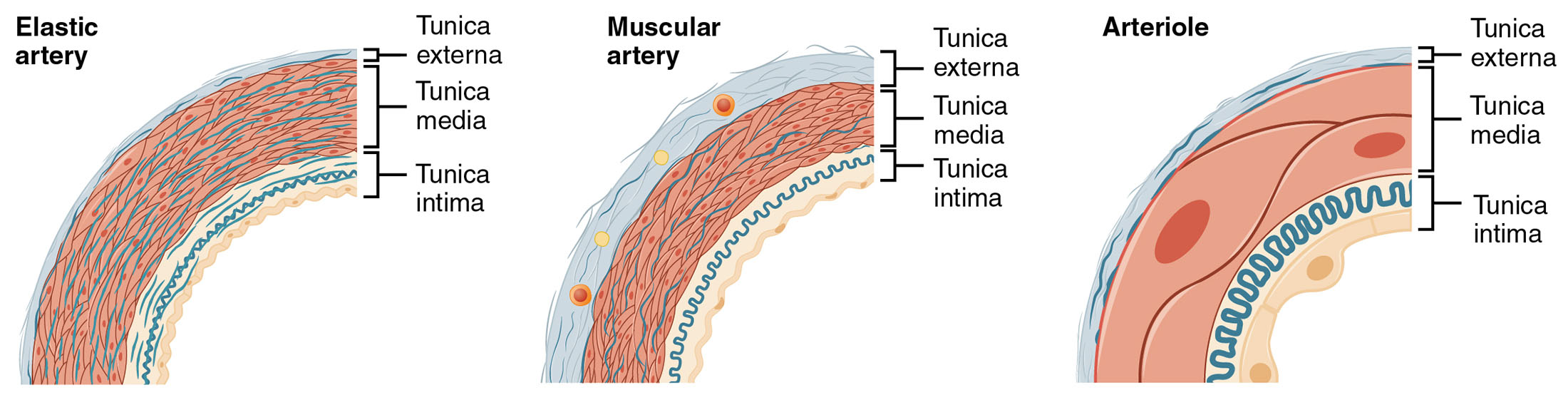
Elastic artery Elastic artery is a large vessel, such as the aorta, characterized by a thick tunica media rich in elastic fibers. These fibers allow the artery to stretch during systole and recoil during diastole, maintaining consistent blood pressure and flow to downstream vessels.
Muscular artery Muscular artery is a medium-sized vessel, like the brachial artery, with a prominent tunica media dominated by smooth muscle cells. This structure enables precise control of blood distribution to specific organs by vasoconstriction or vasodilation in response to metabolic needs.
Arteriole Arteriole is a small vessel that connects arteries to capillaries, featuring a thin tunica media with fewer elastic fibers and more smooth muscle. This design allows arterioles to regulate blood flow into capillary beds, playing a key role in controlling peripheral resistance and blood pressure.
The Role of Arterial Types in Circulation
This overview highlights how each artery type contributes to the circulatory system. Their specialized structures ensure efficient blood delivery tailored to different physiological demands.
- Elastic Artery Function: The extensive elastic lamellae in the tunica media absorb the high pressure of ventricular ejection. This elasticity smooths the pulse wave, protecting smaller vessels.
- Muscular Artery Role: The thick smooth muscle layer responds to neural and hormonal signals, adjusting blood flow to organs like the kidneys or brain. This adaptability supports varying metabolic rates.
- Arteriole Regulation: The narrow lumen and muscular walls control capillary perfusion, influenced by local factors like pH and oxygen levels. This regulation fine-tunes blood distribution.
Anatomical Details of Arterial Types
The image provides a comparative view of arterial structures, emphasizing their layered differences. These distinctions reflect their positions and functions within the vascular tree.
The visual representation aids in understanding the progression from large to small vessels. This knowledge is essential for studying vascular anatomy and physiology.
- Elastic Artery Composition: The tunica media contains up to 40-70 elastic lamellae, interspersed with smooth muscle. The tunica intima features a thick internal elastic lamina.
- Muscular Artery Structure: The tunica media is dominated by circular smooth muscle, with fewer elastic fibers than elastic arteries. The tunica adventitia is thinner, providing minimal support.
- Arteriole Characteristics: The tunica media is reduced to one or two layers of smooth muscle, with a thin tunica adventitia. The endothelial lining is prominent, facilitating flow into capillaries.
- Layer Transitions: The tunica intima remains consistent across types, while the media and adventitia vary in thickness. This gradient supports the transition from high to low pressure.
Physiological Functions of Arterial Types
The physiological roles of these arteries are shaped by their anatomical designs. Their functions ensure effective blood transport and pressure regulation.
Each type plays a distinct role in maintaining circulation. This functionality supports the body’s overall cardiovascular health.
- Elastic Artery Pressure Maintenance: The recoil of elastic fibers during diastole sustains blood pressure, up to 80 mmHg. This mechanism prevents collapse of the vascular system.
- Muscular Artery Distribution: The smooth muscle adjusts lumen size, directing blood to active tissues during exercise. This control is mediated by sympathetic nerves.
- Arteriole Resistance: The narrow diameter creates high resistance, regulating capillary flow. This resistance contributes to about 50% of total peripheral resistance.
- Oxygen Delivery: All types carry oxygenated blood, with elastic arteries distributing it broadly and arterioles fine-tuning delivery. This ensures tissue oxygenation.
Comparative Anatomy Across Arterial Types
The image allows a side-by-side comparison of elastic arteries, muscular arteries, and arterioles. These differences are critical to their specific circulatory roles.
The visual contrast highlights the structural evolution. This understanding is key to grasping vascular dynamics.
- Elastic vs. Muscular Arteries: Elastic arteries have more elastic lamellae, while muscular arteries emphasize smooth muscle. This reflects their roles in pressure maintenance versus distribution.
- Muscular vs. Arterioles: Muscular arteries have a thicker media, while arterioles have a thinner, more contractile layer. This shift supports finer flow control.
- Transition to Capillaries: Arterioles narrow into precapillary sphincters, reducing to a single endothelial layer. This transition maximizes exchange surfaces.
- Histological Features: Elastic arteries show dark elastic bands under staining, while muscular arteries reveal muscle layers. Arterioles display sparse muscle, aiding identification.
Clinical and Research Perspectives
Insights from arterial types inform medical practice and research. Their structures are key to studying cardiovascular health and disease.
Advances in imaging and histology enhance these studies, offering diagnostic tools. These efforts support innovative treatment strategies.
- Atherosclerosis Risk: Plaque buildup in elastic arteries like the aorta narrows the lumen. Microscopic analysis guides statin therapy.
- Hypertension Impact: Thickened tunica media in muscular arteries raises resistance, detectable via imaging. This informs antihypertensive treatment.
- Arteriole Dysfunction: Narrowing in arterioles contributes to hypertension, visible under microscopy. Angiotensin-converting enzyme inhibitors target this.
- Therapeutic Innovations: Stem cell research explores regenerating elastic artery layers. Gene therapy aims to enhance arteriole vasodilation.
In conclusion, this image of arterial types—elastic artery, muscular artery, and arteriole—provides a detailed look at their structural and functional diversity, essential for effective circulation. These anatomical insights not only deepen our understanding of the vascular system but also support advancements in diagnosing and treating cardiovascular conditions.

