Explore the intricate structure of the tooth with this detailed longitudinal section, revealing the relationships between enamel, dentin, and pulp. Learn about the crown, neck, and root, along with supporting structures like the gingiva and periodontal ligament, crucial for comprehensive oral health.
A tooth is a complex and highly specialized organ, far more intricate than its outward appearance suggests. Embedded within the alveolar sockets of the jaw, each tooth is a marvel of biological engineering, designed for durability and a wide range of functions including mastication, speech, and maintaining facial aesthetics. This longitudinal section through a molar provides a clear understanding of its internal and external components, illustrating the vital connections between its hard tissues, soft tissues, and supporting structures that collectively ensure its strength and vitality.
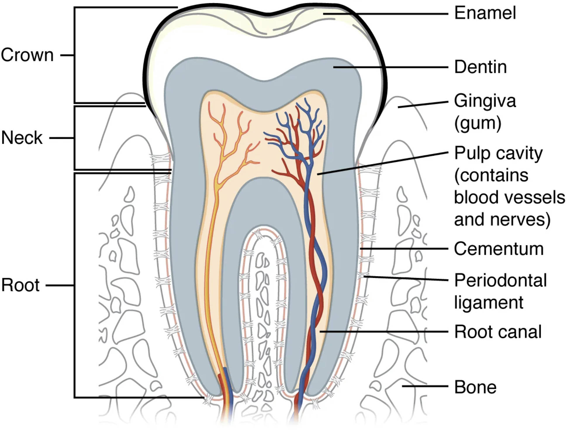
Crown: The crown is the visible part of the tooth that extends above the gum line. It is covered by enamel and is primarily responsible for the mechanical breakdown of food during chewing.
Neck: The neck, also known as the cementoenamel junction, is the constricted area where the crown meets the root of the tooth. This region is typically covered by the gingiva.
Root: The root is the part of the tooth embedded within the alveolar bone of the jaw, below the gum line. It provides anchorage for the tooth and contains the pulp chamber and root canals.
Enamel: Enamel is the outermost, hardest, and most mineralized substance in the human body, covering the crown of the tooth. Its primary function is to protect the underlying dentin and pulp from damage caused by chewing forces and acids.
Dentin: Dentin is a calcified tissue that forms the bulk of the tooth, lying beneath the enamel in the crown and cementum in the root. It is softer than enamel but harder than bone, containing microscopic tubules that transmit sensations like pain and temperature.
Gingiva (gum): The gingiva, or gum, is the soft tissue that surrounds the teeth and covers the alveolar bone. Healthy gingiva provides a protective seal around the tooth, preventing bacteria from entering deeper structures.
Pulp cavity (contains blood vessels and nerves): The pulp cavity is the innermost part of the tooth, extending from the crown (pulp chamber) down into the roots (root canals). It houses the dental pulp, which consists of blood vessels, nerves, and connective tissue, providing vitality to the tooth and sensing stimuli.
Cementum: Cementum is a bone-like tissue that covers the outer surface of the tooth root. It provides an attachment site for the periodontal ligament, securing the tooth in its socket.
Periodontal ligament: The periodontal ligament (PDL) is a specialized connective tissue that surrounds the tooth root and connects it to the alveolar bone. It acts as a shock absorber during chewing and contains sensory nerve endings that provide proprioception.
Root canal: The root canal is a passage within the tooth root that houses the dental pulp, including its blood vessels and nerves. Infections within the root canal often necessitate endodontic treatment.
Bone: The alveolar bone is the part of the jawbone that supports and holds the teeth in their sockets. It provides the foundation for tooth stability and undergoes continuous remodeling throughout life.
The Interconnected Components of a Healthy Tooth
The structure of a human tooth is a testament to biological engineering, meticulously designed to withstand immense forces while maintaining vitality and sensory function. Each component, from the visible crown to the hidden root, plays a specific and crucial role in the overall integrity and health of the tooth. Understanding these interconnected parts is fundamental for appreciating dental health and the importance of preventive care.
The primary components of a tooth can be broadly categorized into:
- Hard Tissues:
- Enamel: The protective outer layer of the crown, incredibly hard and resistant to wear and acid.
- Dentin: The bulk of the tooth, providing structural support and sensory input due to its tubular structure.
- Cementum: Covers the root, anchoring the tooth within the jawbone via the periodontal ligament.
- Soft Tissues:
- Pulp: The living core of the tooth, containing nerves, blood vessels, and connective tissue, responsible for tooth vitality and sensation.
- Supporting Structures:
- Gingiva (Gums): The soft tissue surrounding the tooth, protecting the root and alveolar bone.
- Periodontal Ligament: Connects the cementum to the alveolar bone, absorbing forces and providing proprioception.
- Alveolar Bone: The jawbone that encases the tooth root, providing stable support.
The health of each component is interdependent. For instance, damage to the enamel through decay can expose the underlying dentin, leading to sensitivity and potential progression to the pulp, causing pain and infection. Similarly, inflammation or disease of the gingiva (gingivitis) can advance to periodontitis, affecting the periodontal ligament and alveolar bone, ultimately leading to tooth mobility or loss. Regular dental hygiene practices, including brushing and flossing, are vital for protecting the enamel and gingiva, thereby preserving the entire complex structure. When these protective measures fail, restorative treatments like fillings, root canals, or periodontal therapy become necessary to restore the tooth’s health and function.
In conclusion, the tooth is a sophisticated biological unit, intricately designed for longevity and essential functions within the oral cavity. Its layered structure, from the robust enamel to the sensitive pulp and supportive bone, highlights the importance of comprehensive dental care. A thorough understanding of tooth anatomy empowers individuals to make informed decisions about their oral health, emphasizing the need for proactive prevention and timely intervention to maintain a healthy, functional dentition throughout life.

