Human Lymphatic System: Anatomy, Function and Pathology of Lymph Nodes
The lymphatic system represents one of the most intricate and essential components of human anatomy, serving as the body’s primary defense network against pathogens and maintaining fluid homeostasis throughout tissues. This comprehensive illustration depicts both the macroscopic organization of the lymphatic system throughout the human body and the detailed microscopic structure of a lymph node, including a pathological view showing tumor infiltration. Understanding the lymphatic system is crucial for medical professionals across disciplines, from immunologists studying immune responses to oncologists tracking cancer metastasis. The lymphatic vessels, nodes, and associated organs form a complex network that works in concert with the circulatory system, yet follows distinct anatomical pathways and serves unique physiological functions that are central to immunity, fluid balance, and lipid transport.
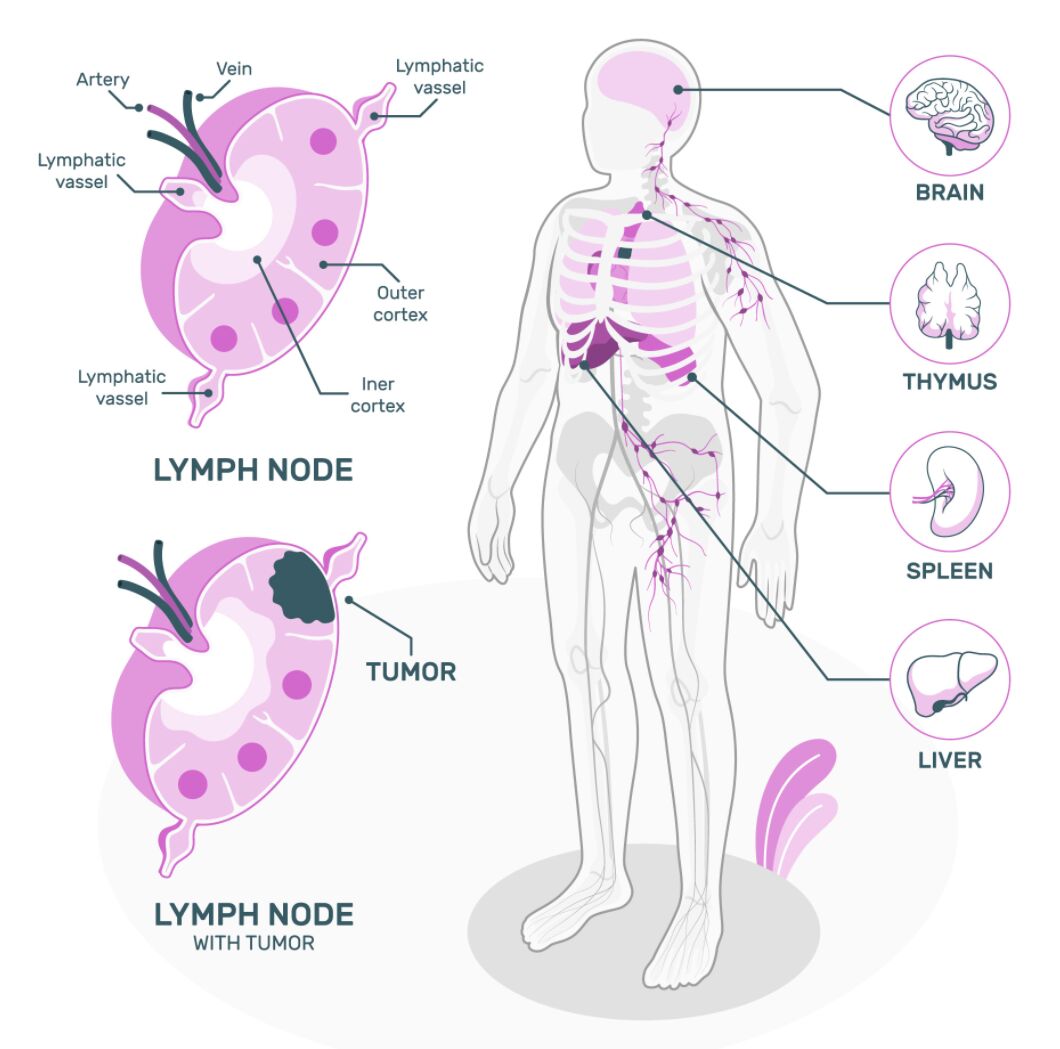
Key Labeled Structures in the Image
Artery: The blood vessel carrying oxygenated blood into the lymph node. These arterial branches supply oxygen and nutrients to the lymphoid tissue within the node and are crucial for maintaining the metabolic activities of the resident immune cells.
Vein: The blood vessel carrying deoxygenated blood away from the lymph node. Veins collect blood after it has circulated through the lymph node’s vascular network and return it to the systemic circulation, completing the blood supply loop to this lymphoid organ.
Lymphatic Vessel: The specialized vascular channels that transport lymph fluid throughout the body. These thin-walled vessels contain valves to prevent backflow and serve as conduits for lymphocytes, antigen-presenting cells, and extracellular fluid collected from tissues.
Outer Cortex: The peripheral region of the lymph node containing primarily B lymphocytes organized into follicles. This region specializes in humoral immunity through the production of antibodies and is the site of B cell activation, proliferation, and germinal center formation following antigen exposure.
Inner Cortex: The deeper region of the lymph node predominantly populated by T lymphocytes. This area facilitates cell-mediated immune responses through T cell interactions with antigen-presenting cells and is crucial for coordinating adaptive immune responses.
Tumor: The abnormal growth of cells within the lymph node representing malignant infiltration. This pathological finding indicates either primary lymphoid malignancy (lymphoma) or metastatic spread from a distant primary tumor through lymphatic drainage pathways.
Brain: The central nervous system organ with emerging relationships to the lymphatic system via recently discovered meningeal lymphatic vessels. The brain’s immune surveillance and waste clearance are partially regulated through connections to the lymphatic system, challenging previous concepts of it being immune-privileged.
Thymus: A primary lymphoid organ located in the anterior superior mediastinum that is crucial for T lymphocyte development and maturation. The thymus is essential for establishing central tolerance and producing a diverse repertoire of T cells capable of recognizing foreign antigens while avoiding autoreactivity.
Spleen: The largest lymphoid organ in the body that filters blood rather than lymph, removing aging erythrocytes and responding to blood-borne pathogens. The spleen contains specialized compartments including white pulp (lymphoid tissue) and red pulp (blood filtration regions).
Liver: A vital metabolic organ with significant immunological functions connected to the lymphatic system. The liver contains a large population of resident macrophages (Kupffer cells) and functions in immune surveillance, contributing to both innate and adaptive immune responses.
The Lymphatic System: Structure and Organization
Anatomical Framework
The lymphatic system represents a sophisticated network that parallels and complements the circulatory system. This complex arrangement of vessels and organs serves multiple crucial physiological functions.
- The lymphatic system consists of lymphatic vessels, lymphoid organs, and lymphoid tissues distributed throughout the body.
- Unlike the circulatory system, the lymphatic system is a unidirectional network that begins as blind-ended capillaries in tissues and progressively joins larger vessels.
- Lymphatic capillaries are highly permeable structures with overlapping endothelial cells that act as one-way valves, allowing interstitial fluid and macromolecules to enter.
- The system progresses from lymphatic capillaries to precollectors, collectors, lymphatic trunks, and finally the thoracic duct and right lymphatic duct that empty into the venous circulation near the subclavian veins.
- Lymphatic vessels contain smooth muscle and one-way valves that prevent backflow, relying on intrinsic contractility, adjacent tissue movement, and respiratory pressure gradients for lymph propulsion.
Lymph Nodes: Microscopic Anatomy
Lymph nodes serve as sophisticated filtration stations strategically positioned throughout the lymphatic network. Their intricate internal architecture optimizes immune surveillance and response.
- Lymph nodes are bean-shaped organs ranging from 1-25mm in size, with approximately 450-700 nodes distributed throughout the human body.
- Each node is encapsulated by connective tissue with trabeculae extending into the parenchyma, providing structural support.
- The node’s internal structure is organized into three distinct regions: cortex, paracortex, and medulla, each with specialized immune functions.
- Afferent lymphatic vessels deliver lymph to the subcapsular sinus, while a single efferent vessel at the hilum allows filtered lymph to exit.
- The cortex contains lymphoid follicles comprising primarily B cells, with germinal centers forming during active immune responses.
- The paracortex (inner cortex) houses predominantly T lymphocytes and dendritic cells, facilitating cell-mediated immunity.
- The medulla contains medullary cords with plasma cells, macrophages, and sinuses that channel lymph toward the efferent lymphatic vessel.
Physiological Functions of the Lymphatic System
Immune Surveillance and Defense
The lymphatic system serves as the body’s principal immunological surveillance network, orchestrating both innate and adaptive immune responses against pathogens. This system provides both immediate protection and immunological memory.
- Lymph nodes continuously filter lymph fluid, allowing resident macrophages and dendritic cells to sample for foreign antigens.
- Upon pathogen detection, antigen-presenting cells process and present antigenic peptides to naive T lymphocytes, initiating the adaptive immune response.
- B lymphocytes in follicles can directly recognize antigens, and with T helper cell assistance, undergo clonal expansion, affinity maturation, and differentiation into antibody-secreting plasma cells.
- The germinal center reaction within lymph node follicles enables somatic hypermutation and class switching, optimizing antibody specificity and function.
- Regional lymphadenopathy (enlarged lymph nodes) often reflects active immune responses occurring within drainage territories, serving as an important clinical sign of infection or malignancy.
Fluid Homeostasis
Beyond immune functions, the lymphatic system plays a critical role in maintaining fluid balance throughout the body’s tissues. This homeostatic mechanism prevents edema and ensures optimal interstitial environments.
- Approximately 20% of the protein-rich fluid that leaks from capillaries into interstitial spaces cannot be directly reabsorbed by venous capillaries due to oncotic pressure dynamics.
- The lymphatic system collects this excess fluid (approximately 3-4 liters daily), preventing interstitial edema and returning it to the bloodstream.
- Initial lymphatics possess specialized junction proteins that create “button-like” connections, allowing interstitial fluid entry while preventing backflow.
- Disruption of lymphatic vessels or nodes, whether through surgery, radiation, infection, or malignancy, can lead to lymphedema—pathological tissue swelling due to impaired lymphatic drainage.
- The lymphatic system’s fluid regulatory capacity is crucial for maintaining tissue pressure and enabling optimal cellular function in all organ systems.
Pathological Conditions of the Lymphatic System
Lymphoid Malignancies
The lymphatic system can itself become the site of primary malignant transformation, giving rise to a diverse group of cancers collectively known as lymphomas. These malignancies originate from lymphocytes at various stages of development.
- Lymphomas represent approximately 3-4% of all malignancies worldwide, with over 80 distinct subtypes recognized in current classifications.
- Hodgkin lymphoma is characterized by the presence of Reed-Sternberg cells within a reactive inflammatory background, typically spreading in a predictable contiguous pattern through lymph nodes.
- Non-Hodgkin lymphomas encompass a heterogeneous group of malignancies varying widely in clinical presentation, cellular origin, and molecular features.
- B-cell lymphomas, including diffuse large B-cell lymphoma, follicular lymphoma, and marginal zone lymphoma, account for approximately 85% of non-Hodgkin lymphomas.
- T-cell lymphomas are generally more aggressive and less common, displaying greater geographic and ethnic variation in incidence.
Metastatic Disease
The lymphatic system frequently serves as a conduit for the spread of cancer cells from primary tumors, representing a critical pathway for metastatic dissemination. Understanding lymphatic drainage patterns is essential for cancer staging and treatment planning.
- Tumor cells can invade lymphatic vessels at the primary site, traveling as emboli through afferent lymphatics to regional nodes, as illustrated in the image showing lymph node with tumor infiltration.
- The concept of sentinel lymph node—the first node receiving drainage from a tumor site—forms the basis for staging procedures in breast cancer, melanoma, and other solid malignancies.
- Patterns of lymphatic spread typically follow predictable anatomical routes, though aberrant drainage can occur, particularly following surgery or radiation that disrupts normal lymphatic channels.
- Extranodal extension, where cancer breaches the lymph node capsule and invades surrounding tissues, indicates more aggressive disease and poorer prognosis in many cancers.
- Modern imaging techniques including lymphoscintigraphy, ultrasonography, and positron emission tomography have enhanced preoperative assessment of lymphatic involvement in malignancy.
Developmental and Age-Related Considerations
Embryological Development
The embryonic origins and developmental dynamics of the lymphatic system provide important insights into its adult structure and function. Understanding these developmental processes helps explain anatomical variations and certain pathological conditions.
- The lymphatic system develops after the cardiovascular system, with the first lymphatic structures appearing around week 5 of human embryonic development.
- Lymphatic endothelial cells differentiate from venous endothelium through the expression of specific transcription factors, particularly Prox1, the master regulator of lymphatic identity.
- Two competing theories explain lymphatic development: the centrifugal theory (sprouting from veins) and the centripetal theory (mesenchymal precursors), with current evidence supporting elements of both models.
- Primary lymphedema can result from genetic mutations affecting lymphangiogenesis during development, leading to hypoplastic or dysfunctional lymphatic vessels.
- The thymus undergoes significant development during fetal life, reaching its maximum relative size at birth and gradually involuting during puberty and adulthood.
Aging and the Lymphatic System
Age-related changes in lymphatic structure and function have important implications for immunity, tissue homeostasis, and disease susceptibility in elderly populations. These alterations contribute to immunosenescence—the decline in immune function with aging.
- The thymus undergoes progressive involution with age, with thymic tissue being gradually replaced by adipose tissue, reducing naive T cell output.
- Lymph node architecture changes with advancing age, including reduction in germinal center formation, alterations in follicular dendritic cell networks, and decreased nodal cellularity.
- Age-related decline in lymphatic vessel density and contractility contributes to less efficient lymph flow and increased susceptibility to lymphedema.
- Impaired lymphatic function with aging may contribute to delayed wound healing, reduced immune surveillance, and altered response to vaccines in elderly individuals.
- Emerging research suggests that interventions to maintain lymphatic vessel integrity may represent a novel approach to mitigating certain aspects of immunosenescence.
Conclusion
The human lymphatic system represents a remarkable biological network that seamlessly integrates several vital functions—immune defense, fluid homeostasis, and macromolecule transport. As illustrated in this comprehensive anatomical representation, the system’s organization spans from microscopic details of lymph node architecture to the macroscopic distribution of lymphatic vessels and organs throughout the body. Understanding the complex structure and function of the lymphatic system is essential for medical professionals across numerous specialties, from immunologists and hematologists to surgeons and oncologists. Recent advances in lymphatic imaging, molecular characterization of lymphatic endothelium, and discovery of novel connections between the lymphatic system and conditions ranging from cancer metastasis to neurological disorders have highlighted the continuing importance of this once-overlooked system. As our understanding continues to evolve, the lymphatic system increasingly reveals itself as not merely an accessory to the cardiovascular system, but a sophisticated network central to health and disease.
- Lymphatic System Anatomy: Comprehensive Guide to Lymph Nodes and Immune Organs
- The Human Lymphatic Network: From Microscopic Lymph Node Structure to Systemic Organization
- Understanding Lymph Node Architecture and the Lymphatic System: A Clinical Perspective
- Lymphatic Vessels and Nodes: Anatomical Framework of the Body’s Defense System
- Lymph Node Microanatomy and the Human Lymphatic System: Essential Knowledge for Medical Professionals

