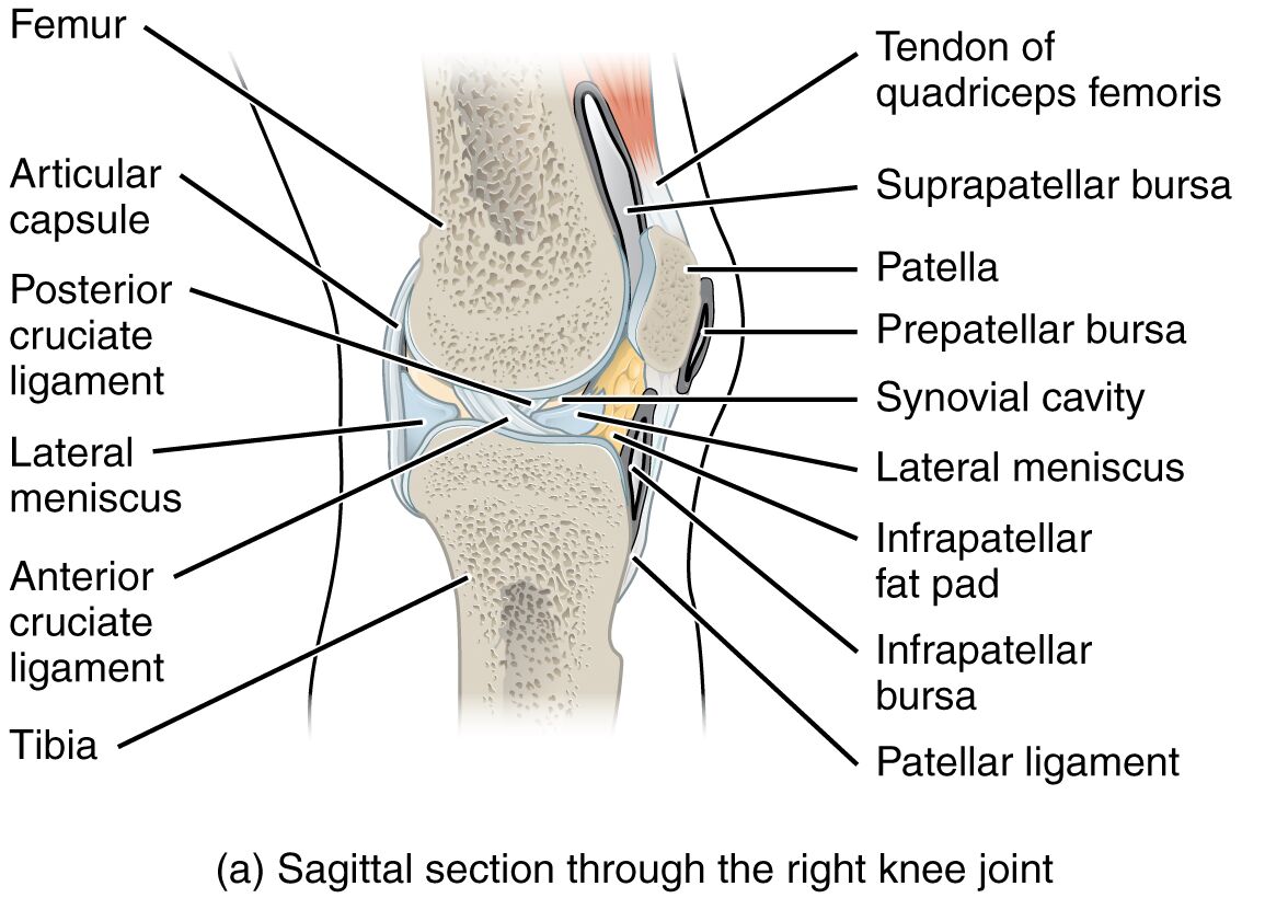
The knee joint, recognized as the largest joint in the human body, plays a crucial role in supporting movement and bearing weight. This sagittal section through the right knee joint provides a detailed view of its complex structure, including bones, ligaments, and bursae, essential for stability and function. Exploring this anatomical image offers valuable insights into the knee’s design and its importance in everyday mobility, making it a key focus for those interested in human anatomy.
Labels Introduction
- Femur: The femur, or thigh bone, forms the upper part of the knee joint and articulates with the tibia to enable leg movement. It provides the primary structural support and strength to the knee, facilitating weight-bearing activities.
- Tendon of quadriceps femoris: This tendon connects the quadriceps muscle to the patella, playing a vital role in knee extension. It ensures powerful leg movements, such as standing up or climbing stairs, by transmitting muscle force.
- Articular capsule: The articular capsule surrounds the knee joint, providing a protective enclosure filled with synovial fluid. It helps maintain joint stability and lubricates the surfaces to reduce friction during movement.
- Suprapatellar bursa: Located above the patella, the suprapatellar bursa reduces friction between the femur and the quadriceps tendon. It acts as a cushion, preventing wear and tear during knee flexion and extension.
- Patella: The patella, or kneecap, is a sesamoid bone that protects the knee joint and enhances the leverage of the quadriceps muscle. It slides within a groove on the femur, aiding in smooth knee motion.
- Prepatellar bursa: Positioned in front of the patella, the prepatellar bursa minimizes friction between the patella and the skin. It is prone to inflammation, known as prepatellar bursitis, due to prolonged kneeling.
- Synovial cavity: The synovial cavity contains synovial fluid, which nourishes the articular cartilage and reduces joint friction. It is a critical component for maintaining the knee’s smooth operation.
- Lateral meniscus: The lateral meniscus is a C-shaped cartilage that acts as a shock absorber between the femur and tibia. It also aids in load distribution and joint stability during movement.
- Infrapatellar fat pad: Located below the patella, the infrapatellar fat pad cushions the knee joint and supports surrounding structures. It can become inflamed, leading to pain during knee flexion.
- Infrapatellar bursa: The infrapatellar bursa lies beneath the patella, reducing friction between the patellar ligament and the tibia. It protects the ligament during knee movement and weight-bearing activities.
- Posterior cruciate ligament: The posterior cruciate ligament (PCL) prevents the tibia from sliding backward relative to the femur. It is essential for maintaining knee stability, especially during activities like walking downhill.
- Anterior cruciate ligament: The anterior cruciate ligament (ACL) restricts forward movement of the tibia and rotational instability. It is a common site of injury in sports, often requiring surgical intervention.
- Tibia: The tibia, or shin bone, forms the lower part of the knee joint and supports the body’s weight. It articulates with the femur, allowing for flexion and extension of the knee.
- Patellar ligament: The patellar ligament connects the patella to the tibia, transmitting the force of the quadriceps muscle. It is crucial for knee extension and is often evaluated in cases of tendonitis.
Detailed Anatomical and Physical Introduction
The knee joint is a marvel of biomechanical engineering, serving as the body’s largest and most complex hinge joint. This sagittal section reveals the intricate interplay of bones, ligaments, and bursae that enable fluid movement and stability. The femur and tibia, as the primary bones, are supported by the patella, which enhances the mechanical advantage of the quadriceps muscle.
Understanding the role of the articular capsule and synovial cavity is key to appreciating how the knee maintains lubrication and nourishment. The menisci, both lateral and medial, play a significant role in shock absorption, protecting the joint from excessive wear. Ligaments such as the ACL and PCL provide critical stability, preventing unnatural movements that could lead to injury.
The bursae, including the suprapatellar, prepatellar, and infrapatellar varieties, act as cushions to reduce friction and protect soft tissues. The infrapatellar fat pad adds an additional layer of protection, while the tendons, like the quadriceps femoris, ensure powerful muscle action. Together, these structures highlight the knee’s adaptability to various physical demands, from walking to athletic performance.
Conclusion
Exploring the sagittal section of the right knee joint offers a deeper appreciation of its anatomical complexity and functional importance. Each component, from the femur to the patellar ligament, contributes to a harmonious system that supports mobility and withstands stress. By understanding these structures, individuals can better recognize the knee’s vulnerabilities and the need for proper care to maintain its health over time.

