Sexual differentiation is a fascinating and complex process that leads to the development of distinct male and female reproductive systems from initially bipotential embryonic structures. This intricate transformation does not commence until the fetal period of development, around week 7 of gestation, highlighting the critical role of genetic and hormonal signals in shaping an individual’s sex. The provided diagram vividly illustrates the key pathways involved, demonstrating how the Wolffian and Müllerian ducts, along with the bipotential gonads, differentiate under the influence of specific cues to form either male or female internal reproductive organs.
Key Structures in Early Sexual Development
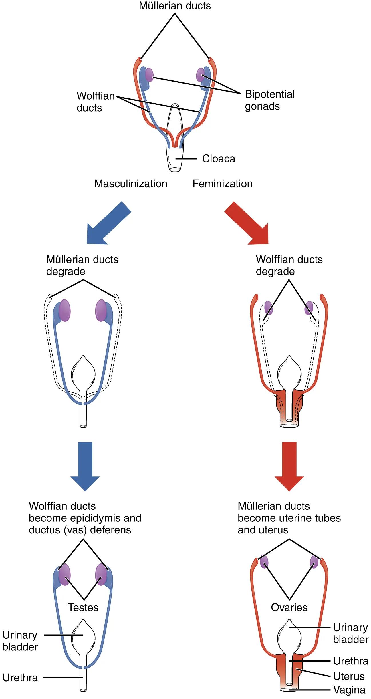
Müllerian ducts: These are paired embryonic ducts that, in the absence of testosterone and Müllerian Inhibiting Substance (MIS), develop into the female internal reproductive organs, including the uterine tubes, uterus, and upper part of the vagina. In males, they typically regress.
Wolffian ducts: Also known as mesonephric ducts, these paired embryonic ducts develop into the male internal reproductive organs in the presence of testosterone, forming the epididymis, vas deferens, and seminal vesicles. In females, they typically regress.
Bipotential gonads: These are the embryonic structures that have the potential to develop into either testes (male) or ovaries (female). Their differentiation is primarily determined by the presence or absence of the SRY gene on the Y chromosome.
Cloaca: A posterior opening that serves as the common exit for the digestive, urinary, and reproductive tracts in early embryonic development. It eventually divides to form separate urogenital and anal openings.
Masculinization: This pathway describes the developmental cascade that leads to the formation of male reproductive structures. It is initiated by the presence of the SRY gene, which directs the bipotential gonads to become testes.
Feminization: This pathway describes the developmental cascade that leads to the formation of female reproductive structures. It occurs in the absence of the SRY gene and the subsequent lack of testosterone and MIS, allowing the Müllerian ducts to develop.
Wolffian ducts degrade: In the absence of testosterone, the Wolffian ducts, which are precursors to male internal genitalia, fail to develop and instead undergo regression.
Müllerian ducts degrade: In the presence of Müllerian Inhibiting Substance (MIS) produced by the Sertoli cells of the developing testes, the Müllerian ducts, which are precursors to female internal genitalia, are suppressed and degrade.
Wolffian ducts become epididymis and ductus (vas) deferens: Under the influence of testosterone, the Wolffian ducts persist and differentiate into the epididymis, which stores sperm, and the vas deferens, which transports sperm.
Testes: The primary male gonads, derived from the bipotential gonads in the presence of the SRY gene. The testes produce testosterone and MIS, crucial for male sexual differentiation.
Urinary bladder: A muscular sac that collects and stores urine before it is excreted. Its development is intertwined with that of the reproductive tracts from the cloaca.
Urethra: The tube that carries urine from the bladder to outside the body. In males, it also carries semen; in females, it is typically shorter and distinct from the reproductive tract.
Müllerian ducts become uterine tubes and uterus: In the absence of MIS, the Müllerian ducts persist and differentiate into the Fallopian tubes (uterine tubes), which transport eggs, and the uterus, where a fertilized egg implants and develops.
Ovaries: The primary female gonads, derived from the bipotential gonads in the absence of the SRY gene. The ovaries produce estrogen and progesterone, critical for female reproductive function.
Vagina: The muscular canal that extends from the uterus to the outside of the body. The upper portion is derived from the Müllerian ducts, while the lower portion forms from the urogenital sinus.
The Hormonal Orchestration of Sex
The initial stages of embryonic development see both male and female embryos possessing a common set of undifferentiated structures: bipotential gonads, Wolffian ducts, Müllerian ducts, and a cloaca. The pivotal moment for sexual differentiation occurs around the seventh week of gestation with the determination of gonadal sex. This is primarily dictated by the presence or absence of the SRY (Sex-determining Region Y) gene on the Y chromosome.
If the SRY gene is present (genetically male, XY), it triggers the bipotential gonads to differentiate into testes. The developing testes then produce two crucial hormones: testosterone and Müllerian Inhibiting Substance (MIS). Testosterone stimulates the Wolffian ducts to develop into the internal male reproductive organs – the epididymis, vas deferens, and seminal vesicles. Simultaneously, MIS causes the Müllerian ducts to regress, preventing the formation of female internal structures.
Conversely, in the absence of the SRY gene (genetically female, XX), the bipotential gonads develop into ovaries. Without testosterone, the Wolffian ducts naturally regress. Furthermore, without MIS, the Müllerian ducts persist and differentiate into the female internal reproductive organs: the uterine tubes, uterus, and the upper portion of the vagina. This default pathway of female development underscores the powerful influence of specific hormonal signals (or their absence) in shaping sexual anatomy.
- Disruptions in this delicate hormonal balance or genetic programming can lead to disorders of sexual development (DSDs), where there is a discrepancy between genetic, gonadal, and anatomical sex.
This intricate process ensures the formation of functionally distinct male and female reproductive systems, essential for the continuation of the species.
Conclusion
The diagram on sexual differentiation provides a clear and concise illustration of how identical embryonic structures diverge into male or female reproductive anatomies. It highlights the critical roles of the SRY gene, testosterone, and Müllerian Inhibiting Substance in orchestrating the development and regression of the Wolffian and Müllerian ducts, ultimately leading to the formation of the testes or ovaries and their associated internal ducts. Understanding this foundational embryological process is vital for comprehending normal reproductive physiology and for diagnosing and managing conditions related to atypical sexual development.

