Staphylococcus aureus, often called golden staph, is one of the most significant bacterial pathogens in human medicine, responsible for a broad spectrum of infections ranging from minor skin boils to life-threatening conditions such as endocarditis and sepsis. This Gram-positive coccus is easily recognized under high-resolution imaging techniques like scanning electron microscopy (SEM), which reveals its characteristic three-dimensional structure and clustering pattern. The provided image offers a stunning close-up view of S. aureus cells, showcasing the classic grape-like arrangement that gives the genus its name and highlighting surface details essential for understanding its biology and pathogenicity.
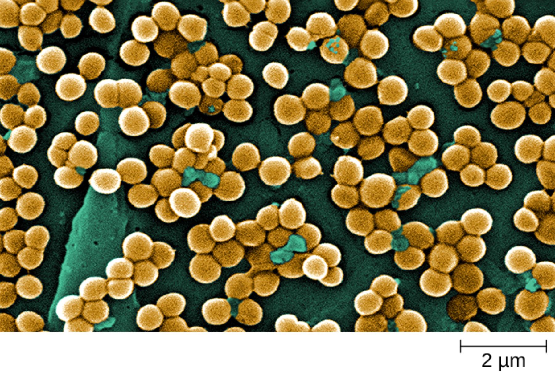
Grape-like clusters refer to the irregular, three-dimensional groupings of spherical bacterial cells visible throughout the SEM image. These clusters form because S. aureus divides successively in multiple planes without complete separation of daughter cells, resulting in the characteristic bunch-of-grapes appearance. In the micrograph, numerous such aggregates of golden-brown cells are seen against the dark background, illustrating how this arrangement enhances adherence and biofilm formation on surfaces.
Spherical cells appear as round, smooth-surfaced cocci approximately 0.5 to 1.5 micrometers in diameter. Each cell in the image displays a uniform, rounded morphology with subtle surface texture typical of staphylococci. The high magnification and 2 μm scale bar allow appreciation of individual cell size and the way cells pack tightly within clusters, a feature that contributes to the organism’s resilience in hostile environments.
2 μm scale bar provides essential context for interpreting the microscopic dimensions shown in the image. Positioned at the bottom, this reference indicates that the field of view encompasses structures on the micrometer scale, confirming the cocci are roughly 1 μm across. Such scaling is critical in electron microscopy to accurately assess cell size, cluster density, and any extracellular material or surface features.
Teal surface highlights represent areas of interest or possible surface proteins, matrix components, or imaging artifacts that appear in contrasting color on some cell clusters. These patches may indicate sites of active division, attachment points, or extracellular polymeric substances. In the context of SEM preparation, they help emphasize three-dimensional topography and potential biofilm-related structures on the bacterial surface.
Morphological Characteristics of Staphylococcus aureus
Under scanning electron microscopy, S. aureus cells exhibit a classic spherical shape with a relatively smooth surface and diameter typically ranging from 0.5 to 1.5 μm. The cells divide in multiple planes, leading to the irregular grape-like clusters prominently displayed in the image. This arrangement distinguishes staphylococci from streptococci, which form chains, and is a key diagnostic feature observed even in routine Gram stains.
The thick peptidoglycan layer in the cell wall contributes to the robust appearance and Gram-positive staining property. In SEM views, cells often appear tightly packed, sometimes with visible septa indicating active division. The image’s high resolution reveals subtle surface irregularities that may correspond to teichoic acids, surface proteins, or early biofilm matrix, all of which play roles in virulence and immune evasion.
- Cells are nonmotile, non-spore-forming, and facultative anaerobes capable of growing in a wide range of conditions.
- Cluster formation enhances protection against desiccation, antibiotics, and host immune responses.
- Colonies on agar plates are typically golden-yellow due to carotenoid pigment production, giving the species its name “aureus.”
Laboratory Identification and Cultural Features
Staphylococcus aureus is readily identified in the laboratory through a combination of microscopic, cultural, and biochemical tests. Gram staining shows Gram-positive cocci in clusters, while SEM, as shown in the image, provides detailed ultrastructural confirmation of the grape-like arrangement. On blood agar, colonies are usually beta-hemolytic with a golden pigment, and the organism tests positive for coagulase, catalase, and mannitol fermentation.
Selective media such as mannitol salt agar exploit the organism’s salt tolerance and ability to ferment mannitol, producing yellow colonies. Advanced techniques like MALDI-TOF mass spectrometry or PCR targeting specific genes offer rapid and accurate identification. The three-dimensional perspective in the SEM image underscores why cluster morphology is so diagnostically valuable even before biochemical confirmation.
- Coagulase production is the hallmark test that differentiates S. aureus from coagulase-negative staphylococci.
- The organism tolerates high salt concentrations and grows well under both aerobic and microaerophilic conditions.
- Modern molecular methods detect methicillin resistance (MRSA) directly from clinical specimens.
Pathogenesis and Virulence Factors
S. aureus possesses an impressive array of virulence factors that enable it to cause diverse infections. Surface proteins such as protein A bind the Fc portion of antibodies, inhibiting phagocytosis, while clumping factor promotes adherence to host tissues and foreign materials. Toxins including alpha-hemolysin, enterotoxins, and toxic shock syndrome toxin-1 contribute to tissue damage and systemic effects.
The grape-like clusters visible in the SEM facilitate biofilm formation on medical devices, wounds, and mucosal surfaces, making infections particularly difficult to eradicate. Biofilms shield bacteria from antibiotics and immune cells, leading to persistent or recurrent disease. The image’s detailed surface view hints at the structural basis for such adherence and community behavior.
- Enzymes like hyaluronidase and lipase aid in tissue invasion and nutrient acquisition.
- Superantigens trigger massive cytokine release, causing conditions like toxic shock syndrome.
- Many strains produce Panton-Valentine leukocidin, associated with severe skin and soft tissue infections.
Clinical Infections and Management
Staphylococcus aureus causes a wide range of infections, from superficial skin lesions such as impetigo, folliculitis, and abscesses to invasive diseases including osteomyelitis, pneumonia, endocarditis, and bacteremia. Skin and soft tissue infections are the most common, often presenting as painful boils or cellulitis. In hospitalized patients, it is a leading cause of surgical site infections and device-related complications.
Treatment depends on the site and severity of infection as well as the strain’s antibiotic susceptibility. Beta-lactam antibiotics remain first-line for methicillin-susceptible strains, while vancomycin or daptomycin are used for methicillin-resistant S. aureus (MRSA). Drainage of abscesses and removal of infected devices are often essential for cure. The SEM image reminds us of the organism’s structural adaptations that complicate therapy.
Prevention strategies include hand hygiene, proper wound care, decolonization protocols for carriers, and infection control measures in healthcare settings. Vaccines targeting surface antigens are under development but not yet widely available. Community-associated MRSA strains have increased the burden of disease outside hospitals, emphasizing the need for ongoing vigilance.
Understanding the ultrastructure revealed by SEM contributes to research on new antimicrobial strategies, including anti-biofilm agents and phage therapy. The grape-like clustering not only aids identification but also explains the organism’s success as a versatile pathogen capable of colonizing humans while remaining ready to cause opportunistic infection.

