In the specialized field of neuroradiology, the visual mapping of blood vessels within the human brain is a vital diagnostic procedure. Arteriography, also known as angiography, provides a high-resolution window into the complex vascular network that sustains our neurological functions. By injecting a contrast medium into the bloodstream and utilizing X-ray technology, clinicians can produce detailed images of the arterial tree, identifying everything from healthy flow patterns to life-threatening obstructions. A normal carotidal arteriography displays the graceful, branching architecture of the vessels as they travel from the neck into the depths of the cranium. Understanding this normal state is the first step for any medical professional in recognizing the subtle deviations that signify disease, such as aneurysms, stenoses, or malformations. This image captures the intricate ‘vascular forest’ that exists within the skull, showcasing the elegance of human circulatory design and the precision of modern medical imaging.
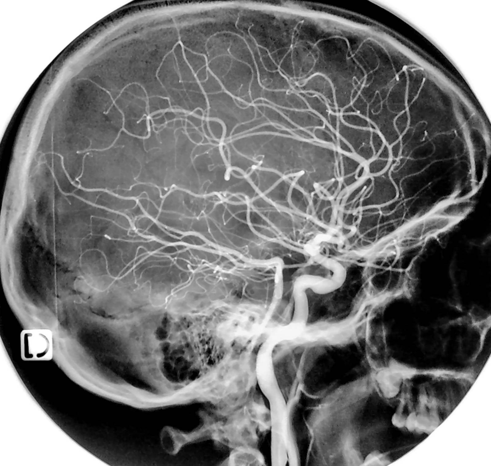
Internal Carotid Artery: This is the large, primary vessel seen ascending through the neck and entering the skull base. It is responsible for providing the vast majority of oxygenated blood to the cerebral hemispheres and is a major focal point in neurovascular studies.
Carotid Siphon: This refers to the distinct S-shaped curvature of the internal carotid artery as it passes through the cavernous sinus near the skull base. This anatomical landmark is highly flexible but can be a common site for the development of atherosclerotic plaques or aneurysms.
Middle Cerebral Artery (MCA): The extensive network of vessels spreading horizontally and backward across the lateral surface of the brain. The MCA is the largest branch of the internal carotid and supplies critical areas involved in motor and sensory function.
Anterior Cerebral Artery (ACA): This branch is seen curving forward and upward toward the midline of the brain’s frontal lobes. It ensures the blood supply to the medial aspects of the cerebral hemispheres, supporting executive function and leg motor control.
Ophthalmic Artery: A relatively small branch visible near the carotid siphon that travels forward toward the eye. Although small on the arteriogram, its presence confirms healthy flow to the visual apparatus and helps orient the radiologist.
Lenticulostriate Arteries: These are fine, hair-like vessels branching off from the proximal segment of the middle cerebral artery. They supply deep structures of the brain like the basal ganglia and are often the site of hypertensive small-vessel strokes.
The Gold Standard of Neurovascular Imaging
While modern medicine has introduced many non-invasive ways to look at the brain, cerebral angiography remains the definitive diagnostic tool for vascular anatomy. The process typically involves a minimally invasive procedure where a catheter is threaded through the femoral artery in the groin up to the carotid arteries in the neck. Once in place, a radio-opaque contrast dye is released, and rapid-fire X-ray images (fluoroscopy) capture the dye as it rushes through the brain. This creates the incredible level of detail seen in the image, where even the smallest capillary-sized branches become visible to the naked eye.
The image provided is a lateral view, which is essential for seeing the depth and vertical orientation of the vessels. Unlike a front-on (AP) view, the lateral view allows radiologists to trace the entire course of the internal carotid artery from its entry into the petrous portion of the temporal bone until it bifurcates into its terminal branches. This view is particularly helpful in identifying the exact location of an aneurysm, which is a weak, bulging spot in an artery wall that could potentially rupture and cause a life-threatening hemorrhage.
Anatomy of the Internal Carotid Artery (ICA)
The ICA is divided into several segments, each with its own clinical significance. As seen in the arteriogram, the vessel enters the skull and makes a sharp, winding turn known as the carotid siphon. This winding path is not just an anatomical quirk; it helps to dampen the high-pressure pulses coming directly from the heart before the blood reaches the delicate tissues of the brain. This segment is closely monitored in patients with cavernous sinus issues or those undergoing complex skull-base surgeries.
Once the ICA emerges into the subarachnoid space, it gives off several important branches. The ophthalmic artery is usually the first major branch, ensuring that the eyes receive a constant supply of blood. Shortly after, the artery splits into its two massive terminal branches: the anterior and middle cerebral arteries. In a normal arteriogram, these vessels should appear smooth, without any narrowings or sudden disappearances of the signal, which would indicate a blockage or a vasospasm.
The Middle Cerebral Artery: The ‘Vascular Forest’
The Middle Cerebral Artery (MCA) is often described as the most important vessel in clinical neurology because it supplies such a vast portion of the brain’s surface. In the lateral arteriogram, you can see the MCA branches spreading out like a fan across the side of the head. These vessels nourish the areas responsible for speech (Broca’s and Wernicke’s areas), hand movement, and facial sensation. Because the MCA territory is so large, a stroke in this artery can lead to significant disability, making its clear visualization on an arteriogram vital for emergency medical decisions.
Radiologists look for the “blush” or “filling” of these branches to ensure that blood is reaching the distal cortex. In cases of an acute ischemic stroke, the arteriogram might show a “cutoff,” where the dye suddenly stops flowing into the MCA forest. This visual evidence allows neurosurgeons to perform a thrombectomy, using a tiny mesh device to physically pull the clot out of the artery and restore the flow of life-sustaining blood to the brain cells.
Clinical Indications for Arteriography
Why would a doctor order a carotidal arteriography? The reasons are diverse and often urgent. One primary indication is the suspicion of an Arteriovenous Malformation (AVM), a tangled mass of abnormal blood vessels where arteries connect directly to veins without a capillary bed. Arteriography is the only way to map the specific “feeder” arteries of an AVM, which is necessary before attempting surgical removal or embolization. Similarly, the procedure is used to evaluate the degree of stenosis (narrowing) in the carotid system, which is a major risk factor for stroke.
- Aneurysm Detection: Providing the highest resolution to plan for clipping or coiling procedures.
- Vasospasm Monitoring: Checking for the narrowing of vessels that can occur after a brain bleed.
- Tumor Mapping: Determining the blood supply of a brain tumor before surgical resection.
- Vascular Dissection: Identifying tears in the arterial wall that can lead to sudden strokes.
Modern Advancements and Patient Safety
While the basic principles of arteriography have remained the same, the technology has advanced significantly. Digital Subtraction Angiography (DSA) is a technique where the computer “subtracts” the image of the skull bones, leaving only the contrast-filled vessels visible against a dark background. This is what gives the image its striking clarity, as the dense bones of the skull are removed to prevent them from obscuring the delicate vascular anatomy. Modern contrast agents are also much safer for the kidneys than those used decades ago.
Despite these advances, the procedure is still invasive and carries a small risk of complications, including bleeding at the puncture site or, rarely, triggering a stroke. Therefore, it is usually reserved for cases where non-invasive tests like MRA (Magnetic Resonance Angiography) or CTA (CT Angiography) do not provide enough detail. For the most complex neurosurgical planning, nothing can replace the raw, real-time data provided by a direct carotidal arteriogram.
Conclusion
The normal carotidal arteriography is a masterpiece of both biological design and medical technology. It reveals a vascular system that is both incredibly complex and highly organized, designed to protect and nourish the human brain under all circumstances. From the powerful trunk of the internal carotid to the fine, wispy branches of the cerebral cortex, every vessel plays a role in our ability to think, move, and experience the world. By studying these images, medical professionals continue to push the boundaries of what is possible in stroke prevention and neurovascular repair, ensuring that the ‘vascular forest’ of the brain remains healthy and vibrant for years to come.

