In the middle of the seventeenth century, the understanding of human anatomy and the natural world was governed by theories that had remained largely unchanged since antiquity. For centuries, medicine relied on the balance of four humors, and the fundamental building blocks of life remained completely invisible to the human eye. The turning point in this narrative occurred in 1665, when a polymath named Robert Hooke published a seminal work that would forever change the trajectory of science. By using a primitive compound microscope to examine a thin sliver of cork, he became the first person to visualize and document the micro-structures of living organisms. These Microscopic Observations of Cork provided the first empirical evidence that large biological structures are composed of smaller, repeating units. While Hooke was looking at the dead cell walls of a plant, his discovery laid the essential groundwork for what we now understand as the cellular basis of all human life, disease, and medical treatment. This article explores the historical significance of Hooke’s work and how his observation of ‘little boxes’ evolved into the foundation of modern pathology and clinical medicine.
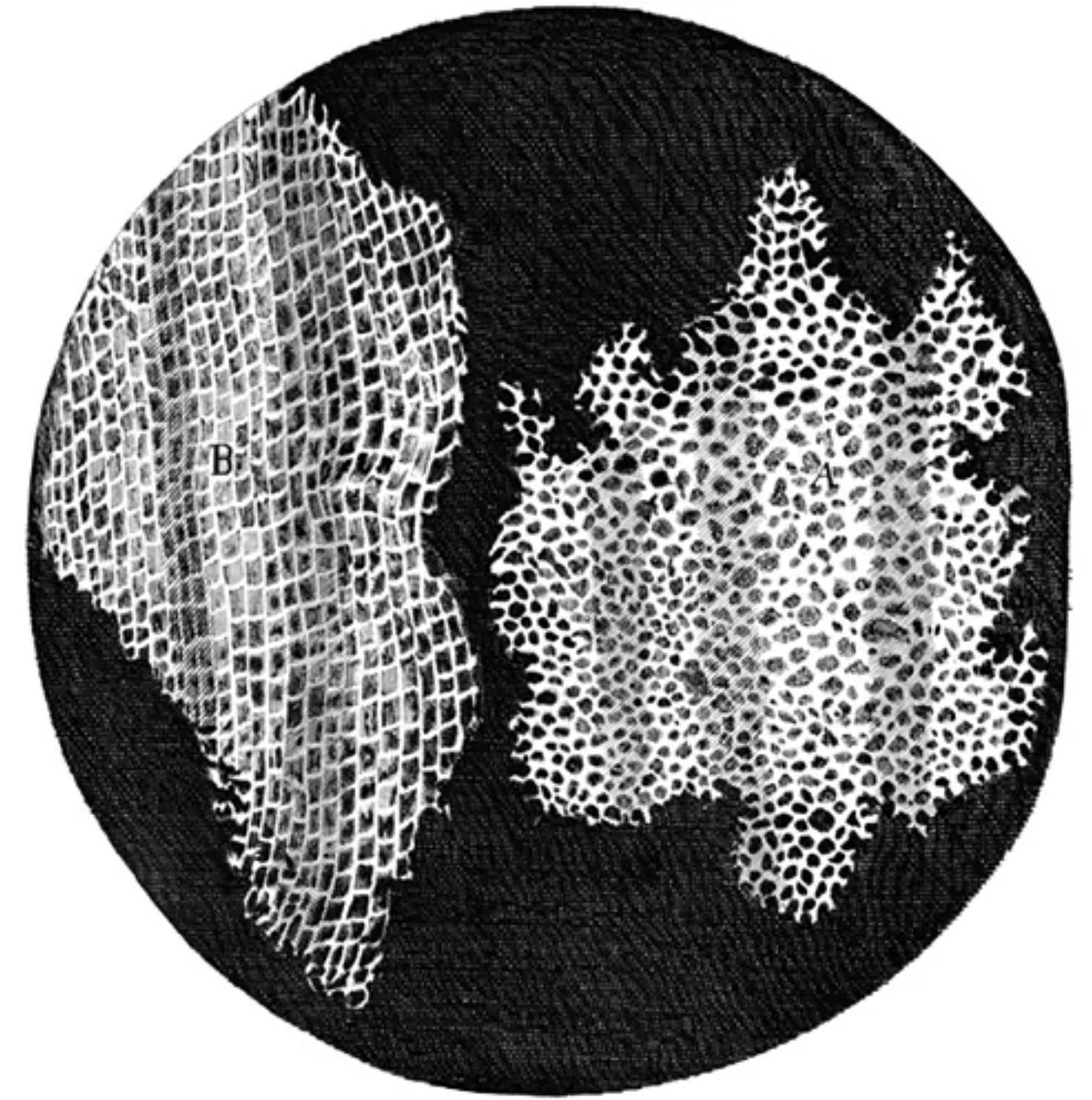
A: This label identifies the transverse or cross-sectional view of the cork tissue, showcasing the distinct honeycomb-like pattern of the cavities. Hooke noted that these pores were remarkably regular and resembled the small rooms, or ‘cella,’ inhabited by monks, leading to the coining of the term ‘cell.’
B: This indicates the longitudinal section of the cork, providing a different perspective on the vertical organization of the cellular structure. From this angle, the repeating box-like units appear more elongated, demonstrating that the structural integrity of the cork is derived from these interconnected microscopic compartments.
The Historical Context of Hooke’s Micrographia
To appreciate the impact of the first Microscopic Observations of Cork, one must understand the intellectual environment of 1665. At the time, the Royal Society of London was a burgeoning hub of the scientific revolution, encouraging scholars to move away from philosophical speculation and toward empirical observation. Hooke’s primary contribution during this period was his masterpiece, Micrographia. This book was not just a scientific report; it was a lavishly illustrated journey into the unseen world. It contained detailed copperplate engravings of everything from the point of a needle to the compound eye of a fly.
Before the publication of this work, the concept of a ‘building block’ for life was non-existent. When Hooke placed a piece of cork under his microscope, he was actually trying to solve a physical mystery: why was cork so buoyant and elastic? By slicing the material extremely thin with a penknife, he was able to see that the solid substance was actually filled with air. The walls he saw were the remains of what we now know as plant cells. While he did not yet understand the biological function of the cytoplasm or the nucleus—which would not be discovered for another 150 years—his ability to categorize and name these structures was the first step toward modern biology.
The Anatomy of Cork and the Naming of the Cell
The choice of cork was particularly fortuitous for the early history of microscopy. Cork is the outer bark of the cork oak tree, *Quercus suber*. As the tissue matures and dies, it leaves behind robust, waxy walls made of suberin. This material is highly resistant to decay and maintains its shape even after the internal living components of the cell have withered away. This meant that Hooke’s primitive lenses, which lacked the clarity and magnification of modern instruments, could still resolve the high-contrast boundaries of these empty chambers.
The term ‘cell’ itself is perhaps the most enduring legacy of Hooke’s Microscopic Observations of Cork. In the 1600s, the word ‘cell’ referred to a small room or a storage compartment. Hooke remarked that the cork looked like a honeycomb, but the individual units reminded him of the sparse living quarters of monks in a monastery. At the time, he did not realize that these ‘cells’ were the universal units of all life. To him, they were unique to the cork and perhaps other plants. However, the nomenclature stuck. Today, whether we are discussing red blood cells, neurons, or cancerous growths, we use the same linguistic root that Hooke established while peering at a piece of bottle stopper.
The Evolution of the Microscope and Observation Techniques
The success of these observations was dependent on the technological advancement of the compound microscope. Hooke used a device featuring two or more lenses, which allowed for greater magnification than simple hand-held magnifying glasses. However, early compound microscopes suffered from significant chromatic and spherical aberrations, meaning the images were often blurry and surrounded by ‘rainbow’ fringes. To compensate for this, Hooke devised an ingenious lighting system using a globe of water to focus light from an oil lamp onto his specimen.
This dedication to the technical aspects of observation is a hallmark of the scientific method. Hooke realized that to see the truth of a structure, he had to master the physics of light. His drawings were so accurate that they remained the gold standard for microscopic illustration for decades. His work encouraged other scientists, such as Antonie van Leeuwenhoek, to refine lens-grinding techniques. Within a few years of Hooke’s discovery, Leeuwenhoek would discover ‘animalcules’—the first living bacteria and protozoa—taking the jump from Hooke’s dead cell walls to the vibrant world of living microbiology.
The Path to Modern Cell Theory
While Hooke’s 1665 observation was the ‘eureka’ moment, it took nearly two centuries for science to fully digest the implications. It wasn’t until the 1830s that German scientists Matthias Schleiden and Theodor Schwann synthesized various observations into a unified cell theory. This theory stated that all living things are composed of cells, that the cell is the basic unit of life, and that all cells arise from pre-existing cells. Hooke’s drawing of cork served as the visual prototype for this entire movement.
In the clinical world, this shift was monumental. Before cell theory, doctors believed diseases were caused by external vapors or spiritual imbalances. Once the cellular nature of life was accepted, medicine became a science of cellular pathology. Scientists like Rudolf Virchow began to realize that a diseased organ was actually a collection of diseased cells. This realization is what allows modern doctors to diagnose cancer by looking for cellular abnormalities in a biopsy or to treat bacterial infections by using antibiotics that specifically target the cellular machinery of the pathogen.
Medical Significance of Cellular Observation Today
Modern medicine is, in every sense, a continuation of Hooke’s Microscopic Observations of Cork. Every time a pathologist looks at a slide under a high-powered electronic microscope, they are following the path blazed by Hooke. The ability to see the ‘little rooms’ of our own bodies allows for the early detection of countless conditions. We can now visualize the internal organelles that Hooke could never have imagined, such as the mitochondria that power our movements or the DNA that contains our blueprints.
- Histology: The study of tissues at the microscopic level is a direct descendant of Hooke’s work, allowing us to see how different cell types organize into functional organs.
- Cytology: This branch of medicine focuses specifically on individual cells to screen for diseases like cervical cancer or leukemia.
- Pharmacology: Drug development relies on understanding how molecules interact with specific receptors on the surface of cell membranes.
- Stem Cell Research: The cutting edge of regenerative medicine involves manipulating the very ‘cells’ that Hooke first named to heal damaged tissues.
The Impact on Public Health and Education
Beyond the lab, Hooke’s discovery had a massive impact on public health. Once it was understood that life—and by extension, germs—operated at a cellular level, the importance of hygiene and sterilization became clear. The visual evidence provided in *Micrographia* made the invisible world ‘real’ for the public. It empowered people to understand that their bodies were not just singular masses, but complex societies of billions of individual units that needed specific care and nutrition.
In medical education, Hooke’s drawing of cork is often the very first image students see. It serves as a reminder that science is built upon the curiosity of those who dare to look closer. It teaches students that even the most mundane object, like a piece of wood or a drop of water, holds secrets that can redefine humanity’s place in the universe. Hooke’s humble observation of cork taught us that the macroscopic world is merely an illusion created by the incredible organization of the microscopic world.
Conclusion: A Legacy That Continues to Grow
The first Microscopic Observations of Cork represent more than just a historical footnote; they are the foundational event of biological science. By peering into the structure of a tree’s bark, Robert Hooke opened a door that led directly to the development of vaccines, the mapping of the human genome, and the creation of life-saving surgeries. His drawings of ‘A’ and ‘B’ in the cork slice remain iconic symbols of the transition from the dark ages of medicine to the era of scientific enlightenment. Today, as we explore the frontiers of nanotechnology and molecular biology, we are still standing on the shoulders of the man who first noticed that life is contained within ‘little boxes.’ Hooke’s legacy is a testament to the power of observation, reminding us that the key to solving the greatest medical mysteries often lies in the smallest details. As long as there are scientists with microscopes and a desire to see the unseen, the journey that began in a slice of cork in 1665 will continue to move toward a deeper understanding of the miracle of life.
Robert Hooke, Microscopic Observations of Cork, Cell Discovery, Micrographia, History of Biology, Cytology, Histology, First Cells, Compound Microscope, Biological Structure

