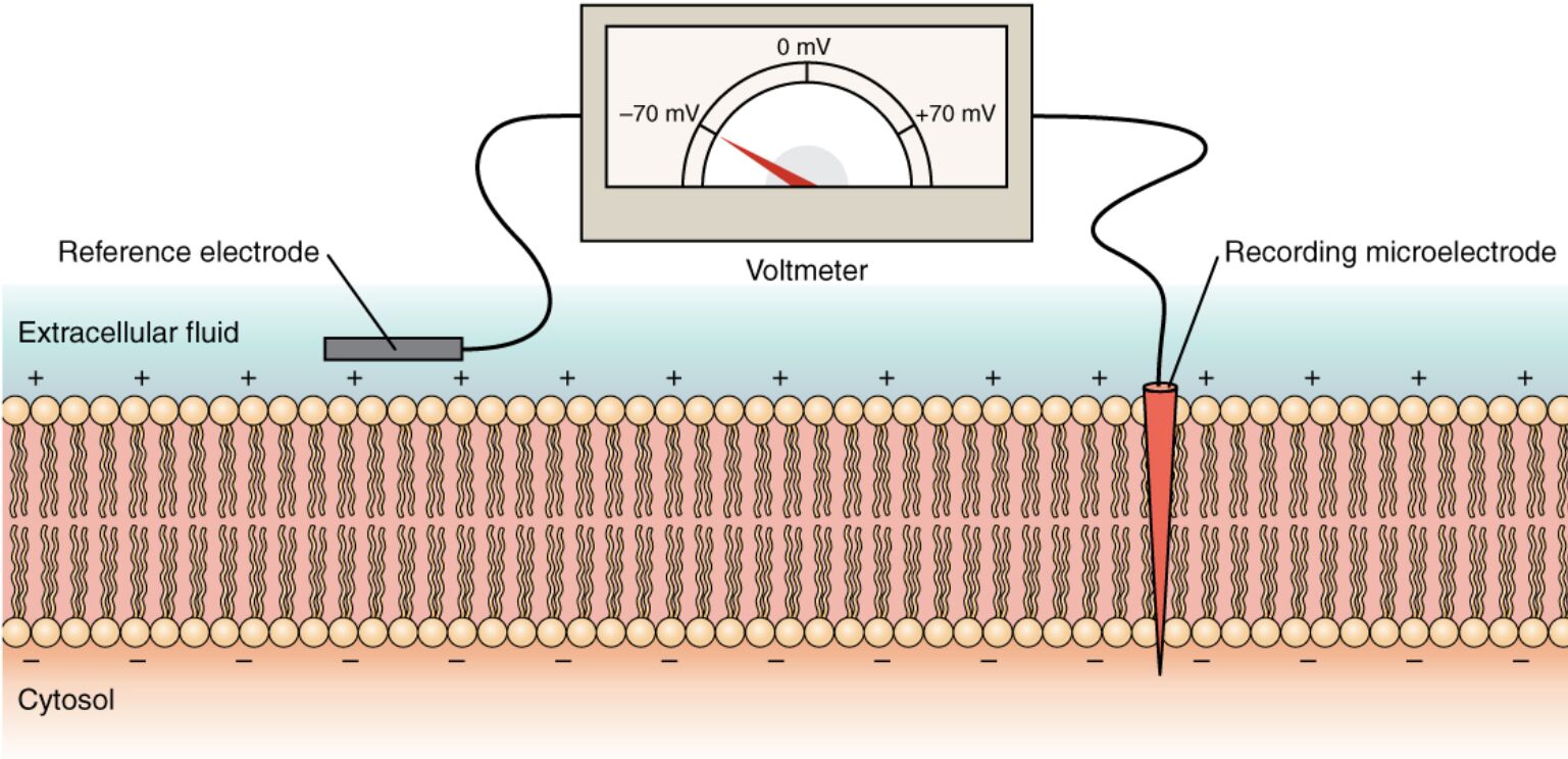
The electrical potential across a cell membrane, known as transmembrane voltage, is a fundamental aspect of cellular function, influencing processes like nerve signaling and muscle contraction. This diagram demonstrates how a recording electrode inside the cell and a reference electrode outside are used with a voltmeter to measure this charge difference, providing a conventional reading relative to the cytosol. Exploring this method offers valuable insights into how scientists and clinicians assess membrane potential and its role in physiological regulation.
Key Labels in the Membrane Charge Measurement Diagram
This section provides detailed explanations for each labeled component, clarifying their roles in measuring transmembrane voltage.
Recording electrode: This fine electrode is inserted into the cell’s cytosol, detecting the electrical potential inside the cell. It transmits this measurement to the voltmeter for analysis of the transmembrane voltage.
Reference electrode: Positioned outside the cell in the extracellular fluid, this electrode provides a baseline potential for comparison. It ensures accurate measurement by stabilizing the external reference point.
Voltmeter: This device compares the electrical potentials detected by the recording and reference electrodes, displaying the transmembrane voltage. It quantifies the difference in charge across the cell membrane.
Cytosol: The internal fluid of the cell where the recording electrode is placed, containing a negative charge relative to the outside. Its potential reflects the activity of ion channels and pumps.
Extracellular fluid: The fluid outside the cell where the reference electrode is located, typically maintaining a positive charge relative to the cytosol. It serves as the external environment for ion exchange.
Cell membrane: The lipid bilayer separating the cytosol from the extracellular fluid, maintaining the charge gradient. It houses ion channels and pumps that regulate the transmembrane voltage.
Transmembrane voltage: The electrical potential difference across the cell membrane, conventionally expressed as negative inside relative to outside. It typically ranges from -70 mV to +30 mV depending on cell type and activity.
Principles of Transmembrane Voltage Measurement
Measuring transmembrane voltage is a cornerstone of cellular physiology. This technique reveals the membrane’s electrical state.
- The recording electrode inside the cytosol detects the internal potential.
- The reference electrode in the extracellular fluid provides the external baseline.
- The voltmeter calculates the difference, yielding transmembrane voltage.
- This method is non-invasive to the cell’s natural function.
- Accurate placement of electrodes is critical for reliable readings.
Role of Electrodes in Voltage Detection
Recording electrode and reference electrode work together to capture potential differences. Their positioning is key.
- The recording electrode penetrates the cell membrane to access the cytosol.
- The reference electrode remains in the extracellular fluid for stability.
- Microelectrodes with high impedance ensure precise measurements.
- Glass pipettes or metal wires are commonly used for these electrodes.
- Calibration adjusts for any electrode drift during experiments.
Function of the Voltmeter in Analysis
The voltmeter translates electrode data into actionable insights. It is essential for quantitative assessment.
- The voltmeter displays the transmembrane voltage in millivolts.
- It amplifies small potential differences for accurate reading.
- Digital voltmeters offer high resolution for dynamic changes.
- The device accounts for electrode resistance and capacitance.
- Real-time monitoring tracks voltage fluctuations during activity.
Structural Context of the Cell Membrane
The cell membrane is the site of electrical activity measurement. Its properties shape the voltage.
- The cell membrane maintains ion gradients via pumps and channels.
- The cytosol and extracellular fluid differ in ion concentrations, like potassium and sodium.
- Lipid bilayers insulate the membrane, enhancing charge separation.
- Proteins within the membrane regulate ion permeability.
- Damage to the membrane can alter transmembrane voltage readings.
Physiological Significance and Clinical Applications
Measuring transmembrane voltage has wide physiological and clinical relevance. It informs health and disease management.
- Transmembrane voltage governs action potentials in neurons and cardiac cells.
- Abnormal voltages are linked to conditions like epilepsy or arrhythmias.
- The voltmeter technique aids in studying channelopathies.
- Intracellular recordings guide pacemaker adjustments in cardiology.
- Research into voltage dynamics supports drug development for nerve disorders.
In conclusion, the measuring charge across a membrane with a voltmeter diagram illustrates how the recording electrode and reference electrode work with the voltmeter to determine transmembrane voltage across the cell membrane. This process, comparing the cytosol to the extracellular fluid, is vital for understanding cellular excitability and signaling. Mastering this technique enhances our ability to explore physiological processes and address related medical challenges effectively.

