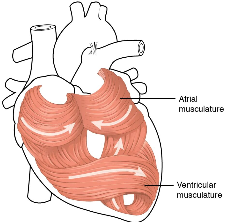
The heart’s ability to pump blood relentlessly relies on its intricate musculature, a marvel of biological engineering. This diagram illustrates the swirling patterns of cardiac muscle tissue, highlighting the atrial musculature and ventricular musculature that drive circulation. Delving into this image reveals the anatomical foundation that supports the heart’s rhythmic contractions and sustains life.
Labelled Parts Explanation
- Atrial musculature The atrial musculature consists of thinner muscle layers in the atria, responsible for contracting to push blood into the ventricles. Its unique arrangement allows for coordinated atrial systole, enhancing ventricular filling.
- Ventricular musculature The ventricular musculature forms the thicker, more robust muscle layers of the ventricles, enabling powerful contractions to pump blood into the pulmonary and systemic circulations. Its spiral and circular fiber orientation maximizes ejection efficiency.
Anatomical Overview of Cardiac Musculature
The heart’s musculature is a complex network that powers its pumping action. This view emphasizes the distinct muscle layers that ensure effective blood flow.
- The atrial musculature supports the atria’s role in receiving and priming blood for ventricular action.
- The ventricular musculature drives the high-pressure ejection of blood, with its thickness varying between the left and right ventricles.
- The swirling pattern of muscle fibers enhances the heart’s twisting motion during contraction.
- This arrangement ensures optimal blood volume is moved with each heartbeat.
The cardiac muscle’s structure is tailored to meet the demands of continuous circulation.
Role of Atrial Musculature in Heart Function
The atrial musculature plays a critical role in the cardiac cycle. Its design supports the initial phase of blood movement.
- The atrial musculature contracts during atrial systole, pushing the final 20-30% of blood into the ventricles.
- Its thinner layers are sufficient for the lower pressure required in the atria.
- The muscle fibers are arranged to facilitate a smooth, wave-like contraction.
- This action is regulated by the sinoatrial node, ensuring timely atrial contribution.
This musculature is essential for maintaining efficient ventricular filling.
Function of Ventricular Musculature
The ventricular musculature is the powerhouse of the heart. Its structure supports the high-pressure demands of circulation.
- The ventricular musculature contracts during ventricular systole, ejecting blood into the aorta and pulmonary trunk.
- The left ventricle’s thicker ventricular musculature generates greater force for systemic circulation, while the right ventricle’s thinner layer suits pulmonary pressure.
- The spiral arrangement of fibers allows a wringing motion, enhancing ejection fraction.
- This design ensures adequate blood supply to both the lungs and the body.
The musculature’s robustness is key to sustaining life-long pumping.
Microscopic Structure of Cardiac Muscle
Cardiac muscle fibers have unique features that support their function. This microscopic view complements the diagram’s macroscopic perspective.
- Cardiac muscle cells are branched and interconnected by intercalated discs, allowing rapid electrical conduction.
- The atrial musculature and ventricular musculature contain sarcomeres, the contractile units that enable muscle shortening.
- Mitochondria are abundant, providing the energy needed for continuous contraction.
- The tissue’s involuntary nature is controlled by the autonomic nervous system and intrinsic pacemakers.
This cellular structure underpins the heart’s endurance and efficiency.
Physiological Significance of Muscle Arrangement
The swirling pattern of cardiac musculature has significant physiological implications. This arrangement optimizes the heart’s mechanical performance.
- The atrial musculature’s orientation supports a gentle squeeze to fill the ventricles.
- The ventricular musculature’s spiral fibers create a twisting motion, improving blood ejection.
- This design reduces energy expenditure while maximizing output.
- The arrangement also aids in distributing mechanical stress evenly across the heart.
Such efficiency is critical for the heart’s lifelong operation.
Clinical Relevance of Cardiac Musculature
Understanding the heart’s musculature is vital for diagnosing and treating cardiac conditions. The muscle layers are key to assessing heart health.
- Thickening of the ventricular musculature can indicate hypertrophic cardiomyopathy, impairing relaxation.
- Damage to the atrial musculature may disrupt atrial kick, affecting ventricular filling.
- Myocardial infarction often involves the ventricular musculature, leading to scar tissue formation.
- Imaging techniques like echocardiography assess muscle thickness and motion.
This knowledge guides therapeutic interventions and improves patient outcomes.
Conclusion
The heart musculature anatomical view provides a detailed look at the muscle layers that drive the heart’s pumping action. By exploring the atrial musculature and ventricular musculature, one gains insight into how their unique arrangements support circulation and cardiac efficiency. This understanding lays a strong foundation for studying cardiovascular physiology and addressing related health concerns, encouraging further exploration of the heart’s remarkable structure and function.

