The spinal cord functions as a vital neural highway, transmitting sensory and motor signals between the brain and the periphery while coordinating reflexes. This cross-sectional view of a thoracic spinal cord segment showcases the organized arrangement of gray matter horns and white matter columns, along with the central canal, essential for processing and relaying information in the central nervous system. This illustration provides a clear depiction of spinal cord architecture, aiding in the understanding of its role in bodily functions.
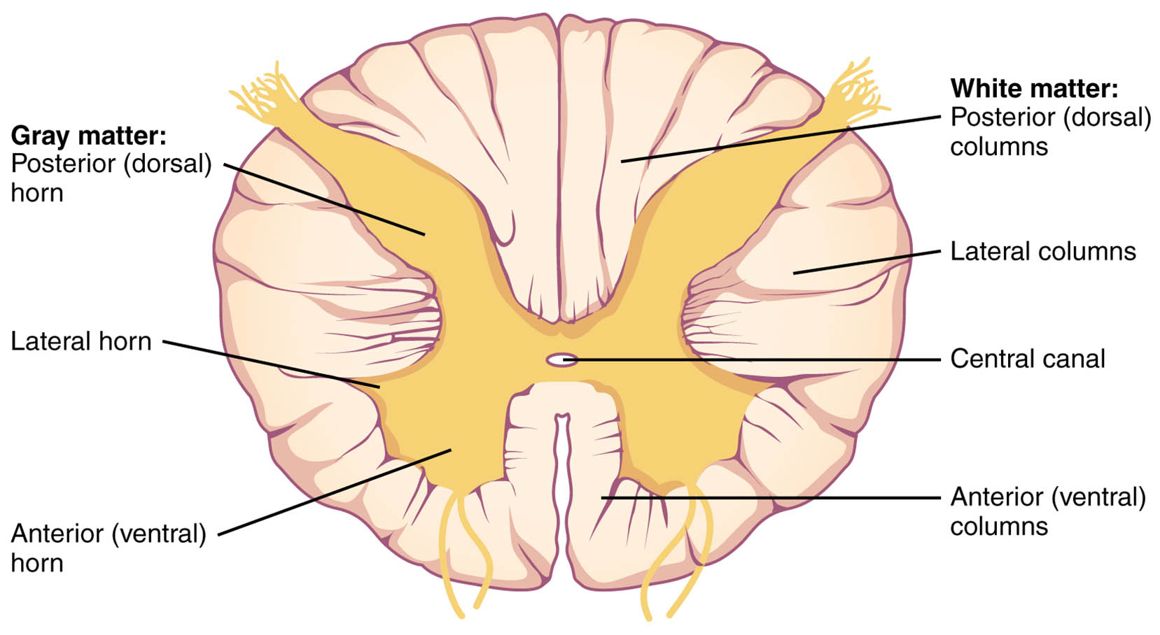
Gray matter: Posterior (dorsal) horn
The posterior (dorsal) horn of gray matter serves as the entry point for sensory afferents from dorsal root ganglia, processing tactile, pain, and temperature signals. It integrates these inputs via interneurons before relaying them to ascending tracts or local circuits for reflex responses.
Lateral horn
The lateral horn, found primarily in thoracic and upper lumbar levels, contains preganglionic sympathetic neurons that regulate autonomic functions such as heart rate and glandular secretion. These neurons extend axons to paravertebral ganglia, forming part of the sympathetic chain essential for fight-or-flight responses.
Anterior (ventral) horn
The anterior (ventral) horn houses alpha and gamma motor neurons responsible for innervating skeletal muscles and maintaining muscle tone. It receives descending inputs from the brain, enabling voluntary movements and participating in spinal reflexes like the stretch reflex.
White matter: Posterior (dorsal) columns
The posterior (dorsal) columns consist of ascending myelinated fibers carrying proprioceptive and fine touch information to the brainstem. These tracts, including the fasciculus gracilis and cuneatus, preserve somatotopic organization for accurate sensory localization in the cortex.
Lateral columns
The lateral columns encompass both ascending spinothalamic tracts for pain and temperature, and descending corticospinal tracts for skilled movements. This region facilitates crossed pathways, supporting contralateral control and integration of sensory-motor functions.
Central canal
The central canal is a narrow, ependyma-lined channel filled with cerebrospinal fluid, extending from the brain’s ventricles. It aids in nutrient diffusion and waste removal, while its surrounding gray commissure allows communication between left and right spinal halves.
Anterior (ventral) columns
The anterior (ventral) columns include descending vestibulospinal and tectospinal tracts that influence posture and head orientation. These fibers also contain some ascending pathways, contributing to overall spinal stability and reflexive adjustments.
Overview of Spinal Cord Anatomy
The spinal cord’s cross-section reveals a symmetrical, H-shaped gray matter core enveloped by white matter, optimized for efficient neural processing. This thoracic view highlights regional specializations unique to autonomic control.
- Gray matter appears butterfly-like, with horns extending into white matter for synaptic integration.
- White matter is divided into three columns per side, each containing specific ascending and descending tracts.
- The cord measures about 42-45 cm in adults, protected by vertebral column and meninges.
- Embryologically, it develops from the neural tube, with gray matter forming from the mantle layer.
Functions of Gray Matter Horns
Gray matter horns serve as sites for synaptic transmission and local circuitry. Their distinct shapes reflect functional divisions in sensory, autonomic, and motor domains.
- Posterior horn processes afferent signals through Rexed laminae, with lamina I handling nociception via substance P release.
- Lateral horn neurons express choline acetyltransferase for acetylcholine synthesis, modulating sympathetic outflow.
- Anterior horn motor pools are organized somatotopically, with flexor muscles medially and extensors laterally.
- Inhibitory interneurons using glycine prevent overexcitation, maintaining reflex balance.
Structure of White Matter Columns
White matter columns facilitate long-distance axonal conduction with myelin insulation. Their organization ensures segregated pathways for different modalities.
- Posterior columns ascend ipsilaterally, synapsing in the medulla before crossing to the thalamus.
- Lateral columns include the spinocerebellar tracts for unconscious proprioception to the cerebellum.
- Anterior columns support the anterior corticospinal tract, primarily for axial muscle control.
- Oligodendrocytes wrap axons in myelin, enabling saltatory conduction at nodes of Ranvier.
Role of the Central Canal
The central canal maintains the spinal cord’s internal milieu. Its presence echoes the cord’s ventricular origins.
- Ependymal cells secrete cerebrospinal fluid components, supporting metabolic exchange.
- In some adults, the canal may narrow or obliterate, but remains patent in youth.
- Surrounding subependymal zone harbors neural stem cells, potential for regeneration.
- Fluid dynamics aid in pressure equalization during movement.
Physiological Integration and Pathways
The spinal cord integrates peripheral inputs with central commands for coordinated responses. Gray and white matter collaborate in reflex arcs and voluntary actions.
- Sensory neurons enter via dorsal roots, synapsing in posterior horns before ascending in white matter.
- Motor outputs exit ventral roots, influenced by descending tracts in lateral and anterior columns.
- Autonomic reflexes from lateral horns bypass higher centers for rapid visceral adjustments.
- Segmental levels vary, with thoracic emphasizing sympathetic innervation.
Clinical and Functional Significance
Understanding spinal cord anatomy informs neurological assessments and interventions. Its vulnerability highlights the need for protective mechanisms.
- Imaging like MRI visualizes cross-sections, detecting lesions in specific columns.
- Transections disrupt pathways, causing sensory loss below the level in posterior columns.
- Autonomic dysreflexia arises from lateral horn dysregulation in injuries.
- Reflex testing evaluates anterior horn integrity through myotatic responses.
Conclusion
The cross-section of the spinal cord exemplifies the organ’s precise design for neural efficiency, with gray matter horns processing local signals and white matter columns enabling transmission. This thoracic perspective, featuring the lateral horn and central canal, underscores its multifaceted role in sensation, movement, and autonomic balance. Exploring these structures deepens appreciation for the nervous system’s complexity and supports advancements in spinal health.

