The lateral view of the human skull provides essential insights into the complex arrangement of bones, sutures, and anatomical landmarks critical for medical diagnosis and surgical planning. This perspective reveals key structures involved in cranial development, sensory function, and mastication.
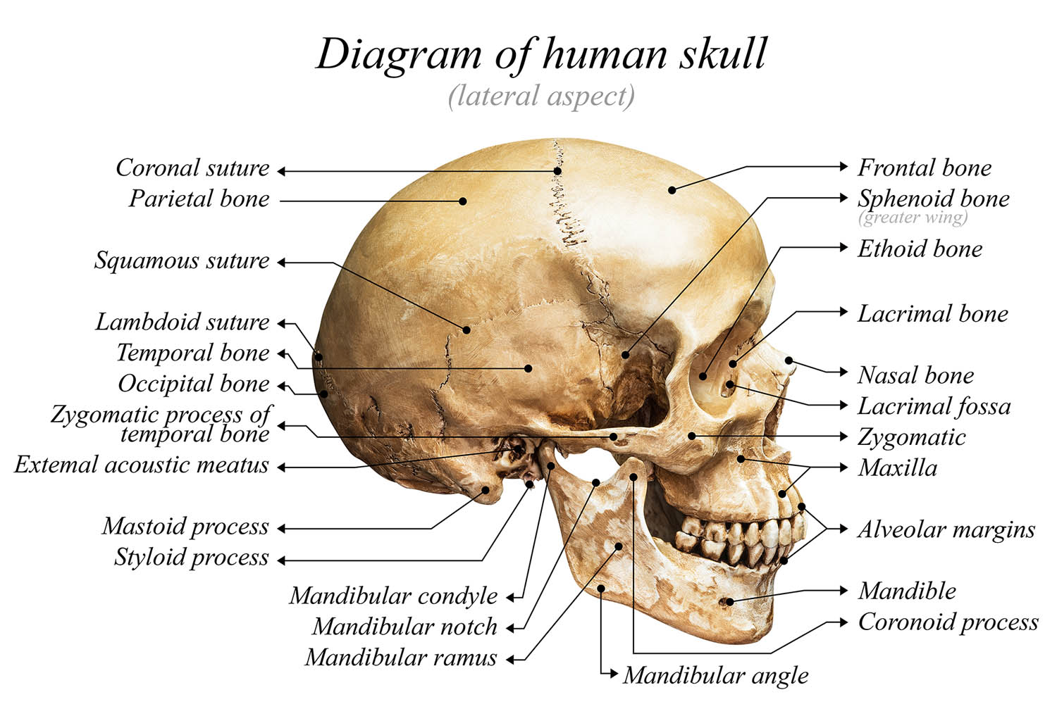
Coronal suture A major cranial suture joining the frontal and parietal bones. This fibrous joint allows for minor movement during birth and early development.
Frontal bone Forms the forehead and anterior cranial vault. Contains the frontal sinuses and provides protection for the frontal lobes.
Sphenoid bone (greater wing) A complex butterfly-shaped bone forming part of the cranial floor. Houses important foramina for neurovascular structures.
Ethoid bone Forms part of the nasal cavity and orbital walls. Contains the cribriform plate for olfactory nerve transmission.
Parietal bone A paired bone forming the superior and lateral aspects of the skull vault. Contains crucial attachment points for temporal muscles.
Squamous suture Joins the temporal and parietal bones. Characterized by its overlapping scale-like appearance.
Lambdoid suture Connects the occipital bone with the parietal bones. Named for its resemblance to the Greek letter lambda.
Temporal bone Houses the middle and inner ear structures. Contains vital pathways for cranial nerves VII and VIII.
Occipital bone Forms the posterior and inferior portions of the skull. Contains the foramen magnum for spinal cord passage.
Zygomatic process of temporal bone Projects anteriorly to join the zygomatic bone. Forms part of the zygomatic arch for muscle attachment.
External acoustic meatus The external opening of the ear canal. Conducts sound waves to the tympanic membrane.
Mastoid process A prominent projection of the temporal bone. Serves as an attachment site for neck muscles.
Styloid process A long, thin projection from the temporal bone. Provides attachment for the stylohyoid ligament and muscles.
Mandibular condyle The articulating surface of the mandible. Forms the temporomandibular joint with the temporal bone.
Mandibular notch A concave area between the condylar and coronoid processes. Allows passage of masseteric vessels and nerve.
Mandibular ramus The vertical portion of the mandible. Provides attachment for muscles of mastication.
Mandibular angle The junction between the body and ramus of the mandible. Shows significant variation in morphology between individuals.
Coronoid process A thin, triangular projection of the mandible. Serves as the attachment site for the temporal muscle.
Alveolar margins The tooth-bearing portion of the maxilla and mandible. Contains individual sockets for dental roots.
Mandible The largest and strongest facial bone. Forms the lower jaw and houses the lower dentition.
Cranial Development and Structural Organization
The human skull’s development follows complex patterns of ossification and suture formation. The lateral view demonstrates the integration of both neural and facial components through distinct growth centers.
Embryological Origins and Growth
The cranial bones arise from neural crest cells and mesoderm. This developmental process involves both intramembranous and endochondral ossification.
Clinical Significance of Lateral Skull Anatomy
Important Surgical Landmarks
The lateral skull view provides critical reference points for neurosurgical approaches. Major vessels and nerves follow predictable paths in relation to these landmarks.
Radiological Assessment
Modern imaging techniques require thorough knowledge of lateral skull anatomy. CT and MRI interpretations depend on understanding normal anatomical relationships.
Temporomandibular Joint Complex
The TMJ represents a unique articular system allowing both hinge and sliding movements. Its complex anatomy includes:
- Articular disc
- Synovial compartments
- Ligamentous attachments
- Muscular components
Functional Considerations
The temporomandibular joint’s biomechanics involve:
- Rotation during initial opening
- Translation during wide opening
- Complex muscle coordination
- Disc movement patterns
Neurovascular Relationships
Cranial Nerve Pathways
Multiple cranial nerves traverse the lateral skull region:
- Facial nerve through temporal bone
- Trigeminal nerve branches
- Vestibulocochlear nerve path
Vascular Supply
Critical vessels course through the lateral skull:
- Middle meningeal artery
- Superficial temporal artery
- Maxillary artery branches
Clinical Applications and Pathology
Common Disorders
Lateral skull pathologies include:
- Temporal bone fractures
- TMJ disorders
- Mastoid infections
- Skull base tumors
Diagnostic Approaches
Modern assessment techniques include:
- Advanced imaging protocols
- Clinical correlation
- Functional testing
- Dynamic assessment
- “Lateral Skull Anatomy: A Comprehensive Guide for Medical Professionals”
- “Understanding Human Skull Anatomy: Lateral View Perspectives”
- “Clinical Analysis of Lateral Skull Features: Expert Review”
- “Advanced Guide to Lateral Cranial Anatomy”
- “Surgical Anatomy of the Lateral Skull: Professional Reference”

