The human foot is a complex mechanical structure consisting of 26 bones, 33 joints, and over a hundred muscles, tendons, and ligaments. This intricate network is designed to perform dual functions: acting as a mobile adapter to navigate uneven terrain and a rigid lever to propel the body forward during locomotion. For medical students and clinicians, mastering the skeletal anatomy of the foot is essential for interpreting radiographic images and diagnosing a wide range of musculoskeletal conditions, from acute trauma to chronic degenerative diseases.
Label-by-Label Explanation
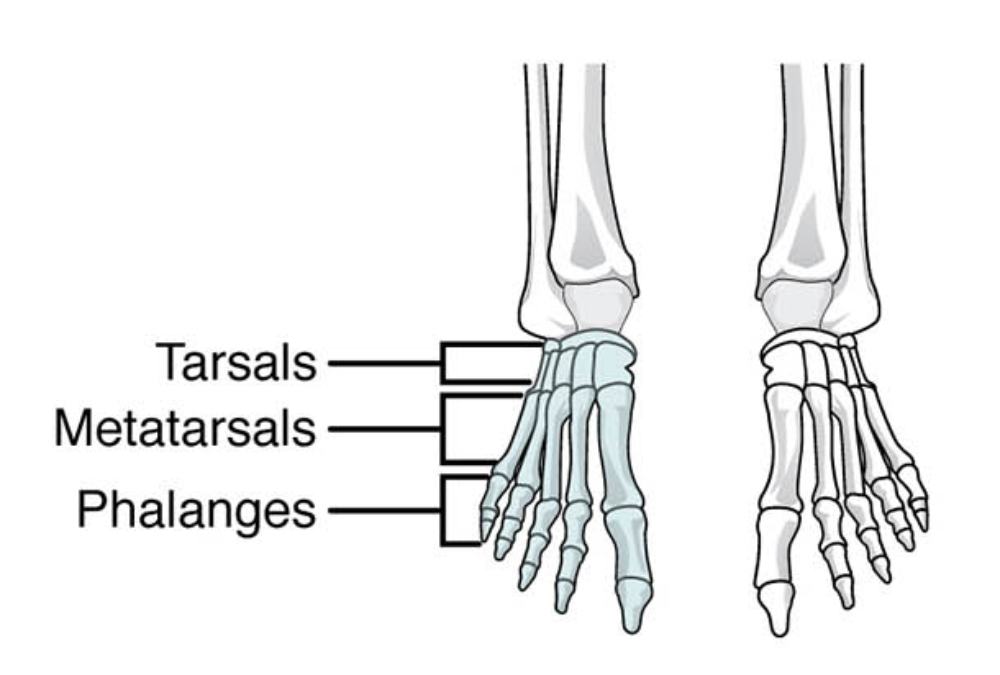
Tarsals
The tarsals are a group of seven short bones that make up the posterior and mid-regions of the foot. These include the talus, calcaneus, navicular, cuboid, and the three cuneiform bones, which together provide the structural foundation for the arches of the foot. Clinically, this region is vital for weight-bearing and is a common site for pathologies such as tarsal tunnel syndrome or osteochondral lesions of the talus.
Metatarsals
The metatarsals are five long bones located between the tarsals and the phalanges, numbered one through five starting from the medial side (great toe). They form the primary structure of the forefoot and are essential for distributing body weight during the different phases of gait. Common clinical issues related to these bones include stress fractures, often seen in athletes, and Lisfranc injuries involving the tarsometatarsal joint complex.
Phalanges
The phalanges are the small bones that comprise the toes, with fourteen in total across each foot. The hallux (great toe) contains only two phalanges (proximal and distal), while the lateral four toes typically contain three segments: proximal, middle, and distal phalanges. These bones provide the necessary length and leverage for balance and propulsion, and they are frequently subject to fractures or deformities like hammer toes.
Anatomical Overview
From an educational perspective, the foot is traditionally divided into three functional segments: the hindfoot, the midfoot, and the forefoot. The hindfoot consists of the talus and the calcaneus (heel bone), which are primarily responsible for early weight-bearing and forming the ankle joint with the tibia and fibula. The midfoot acts as a shock-absorbing bridge, containing the navicular, cuboid, and cuneiforms. Finally, the forefoot encompasses the metatarsals and phalanges, which provide the platform for terminal stance and toe-off.
The skeletal arrangement visible in this anterior (dorsal) view highlights the longitudinal organization of the foot. The articulation between the tarsals and metatarsals, known as the tarsometatarsal joints, is a critical structural transition zone. The second metatarsal is particularly noteworthy as it is often the longest and most rigid, acting as a cornerstone that stabilizes the midfoot-forefoot junction.
Functional Significance
The bones of the foot do not sit in a flat plane; rather, they are arranged to form three distinct arches: the medial longitudinal, lateral longitudinal, and transverse arches. These arches are maintained by the interlocking shapes of the bones and reinforced by strong ligaments like the plantar fascia. The tarsals, specifically the talus, act as the keystone of the medial arch, receiving the full weight of the body and distributing it posteriorly to the calcaneus and anteriorly through the metatarsal heads.
During ambulation, the foot must transition from a flexible “bag of bones” during heel strike (to absorb impact) to a rigid lever during toe-off. This transition is mediated largely by the subtalar joint and the midtarsal joints. If this mechanical synchronization is disrupted due to bony malalignment or ligamentous laxity, it can lead to conditions such as pes planus (flat feet) or pes cavus (high arches), both of which significantly alter gait biomechanics.
Clinical Relevance
Understanding the spatial relationships of these bones is paramount in clinical practice. For instance, the base of the fifth metatarsal is a frequent site of Jones fractures or avulsion fractures due to the pull of the peroneus brevis tendon. In the phalangeal region, the first metatarsophalangeal (MTP) joint is the most common site for gout (podagra) and hallux valgus (bunions), both of which cause significant pain and mobility impairment.
In patients with systemic diseases like diabetes, the bones of the midfoot and tarsals are at risk for Charcot foot, a progressive condition characterized by bone fragmentation and joint collapse. Early recognition of structural changes in the metatarsal and tarsal regions can prevent catastrophic foot deformities and ulcers. Furthermore, in trauma surgery, the precise reduction of the metatarsal shafts is critical to ensuring a normal weight-bearing pattern and avoiding long-term metatarsalgia.
Diagnostic or Educational Importance
For healthcare learners, the skeletal anatomy shown in this image serves as the roadmap for physical examination and radiological interpretation. Identifying the “tarsal-metatarsal-phalangeal” progression allows clinicians to localize pathology accurately. For example, pain localized to the tarsal region may suggest a stress fracture of the navicular, while pain in the phalanges often points toward trauma or digital deformities.
Radiologists utilize standard views—Anteroposterior (AP), Oblique, and Lateral—to assess these structures. On an AP view, like the one represented in this illustration, one would look for the alignment of the medial borders of the second metatarsal and the intermediate cuneiform to rule out Lisfranc ligamentous injuries. Mastery of these basic anatomical divisions is the prerequisite for understanding more complex orthopedic concepts and surgical approaches to the lower extremity.
In summary, the foot’s skeletal framework is a sophisticated system designed for endurance and precision. By categorizing the bones into tarsals, metatarsals, and phalanges, medical professionals can better understand the biomechanical requirements of the foot and provide more accurate diagnoses for their patients. Practical learning involves correlating these anatomical landmarks with palpable structures on a live patient to bridge the gap between textbook knowledge and clinical application.
Medical Learning Tips
- The hallux (great toe) is unique among digits because it lacks a middle phalanx.
- The second metatarsal base is the most stable and least mobile of the metatarsals, often making it the pivot point for the foot.
- The tarsal bones, particularly the talus, are the only structures that articulate directly with the leg bones to form the ankle joint.

