Eukaryotic cells represent one of the most diverse domains of life, exhibiting a vast array of shapes and sizes that are intricately tied to their ecological niches and physiological requirements. The study of spheroid organisms, such as the Chromulina alga, provides a window into how complex internal architectures are packed into microscopic volumes. By examining these single-celled eukaryotes, we gain a better understanding of the fundamental principles of cellular anatomy, motility, and metabolic efficiency that sustain life across the planet’s diverse ecosystems.
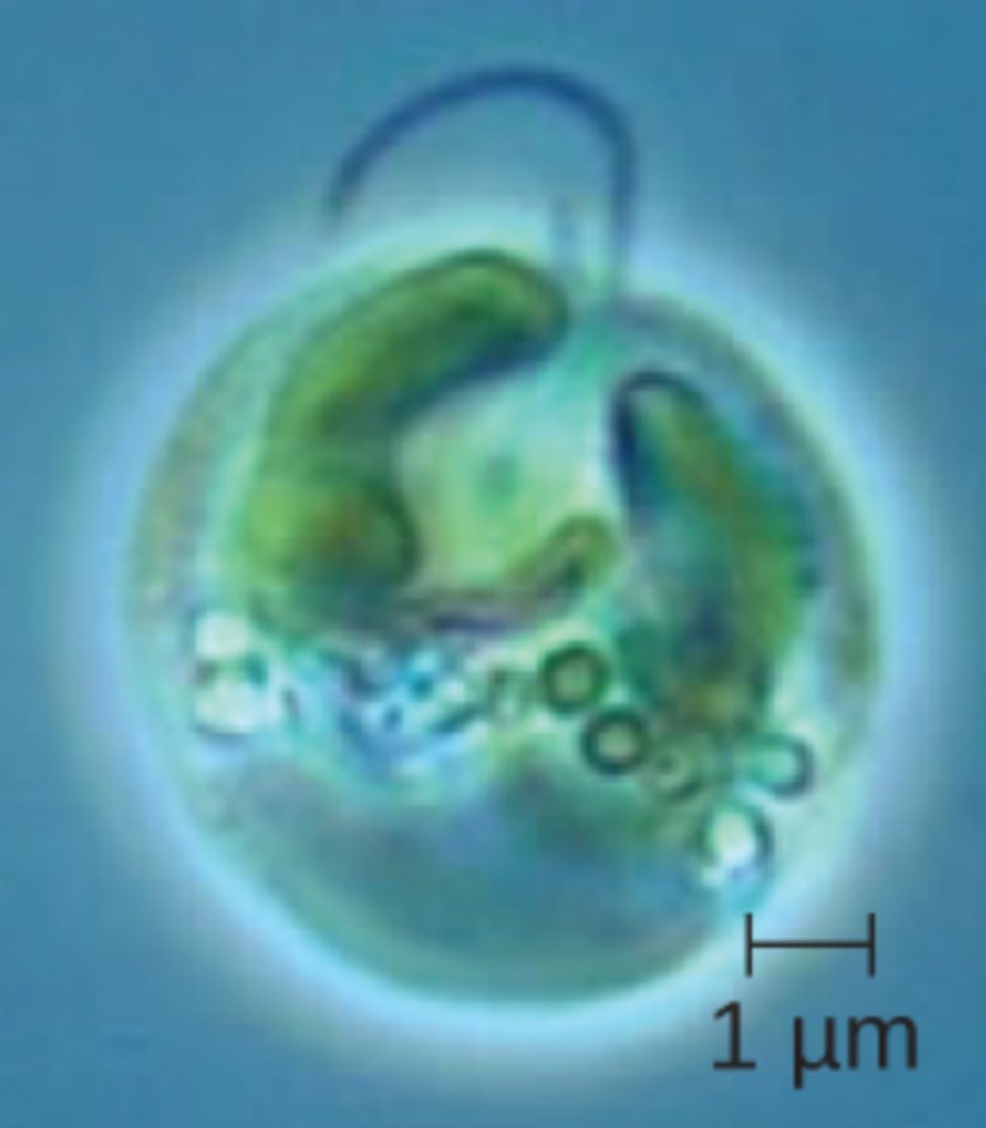
1 μm: This scale bar indicates a measurement of one micrometer, which is equal to one-millionth of a meter. It provides a crucial reference for the observer, demonstrating the extremely small size of this complex eukaryotic organism.
Eukaryotic Cell Diversity: Analyzing the Anatomy of Spheroid Chromulina Algae
The world of eukaryotic biology is characterized by an incredible degree of morphological variation, ranging from the elongated neurons of the human nervous system to the perfectly rounded shapes of certain aquatic protists. This variation is not random; rather, it is a direct result of evolutionary adaptations that allow cells to optimize their interactions with the surrounding environment. In unicellular eukaryotes, the cell’s exterior shape is often a compromise between the need for efficient nutrient uptake and the mechanical requirements for movement.
A primary distinguishing feature of these organisms is the presence of membrane-bound organelles, which allow for the compartmentalization of specialized biochemical reactions. In the case of Chromulina, a genus of golden-brown algae, the cell contains distinct structures for photosynthesis, energy production, and waste management. These internal compartments enable the cell to perform high-level functions that simpler prokaryotic cells cannot achieve at the same scale.
Key characteristics of eukaryotic cell morphology include:
- Variable surface-to-volume ratios that influence the rate of diffusion and nutrient absorption.
- Specialized appendages, such as flagella or cilia, used for locomotion and sensory perception.
- Dynamic internal cytoskeletons that maintain cell shape and facilitate the transport of vesicles.
- Diverse protective outer layers, ranging from flexible plasma membranes to rigid cell walls.
Understanding the structural blueprint of these cells is essential for fields ranging from environmental microbiology to evolutionary medicine. By analyzing how a spheroid cell like Chromulina manages its internal resources, researchers can draw parallels to the functioning of specialized cells in multicellular organisms. This microscopic perspective is fundamental to our comprehension of the biological “building blocks” that form the basis of all complex life.
The Spheroid Architecture of Chromulina Algae
The Chromulina alga depicted in the micrograph serves as a classic example of a spheroid eukaryotic cell. Anatomically, this shape is highly efficient for organisms living in fluid environments where they must maintain buoyancy. The rounded form minimizes the surface area exposed to the environment relative to its internal volume, which can be a protective adaptation against certain types of environmental stress or predation.
Within the cytoplasm of this alga, one can observe large, pigmented organelles known as chloroplasts. These structures are the site of photosynthesis, where light energy is converted into chemical energy in the form of glucose. The physiological process of capturing photons requires a high degree of organization, with internal thylakoid membranes stacked to maximize the surface area for light-harvesting pigments. In Chromulina, these chloroplasts often take on a lobed or C-shaped appearance, wrapping around other internal structures like the nucleus.
Motility and Sensory Physiology
One of the most striking features of many Chromulina species is their ability to move through the water column. This is typically achieved through flagellar movement, involving a long, whip-like appendage that protrudes from the cell body. The flagellum is powered by dynein motor proteins that slide microtubules against one another, creating a rhythmic beating motion. This allow the alga to move toward light sources (phototaxis) or nutrient-rich areas (chemotaxis), which is vital for its survival as a primary producer in aquatic food webs.
In addition to motility, the cell must maintain an active osmotic balance with its surrounding environment. Because freshwater environments are often hypotonic compared to the cell’s interior, water constantly enters the cell via osmosis. To prevent the cell from bursting, many eukaryotes utilize contractile vacuoles that act as bilge pumps, collecting excess water and periodically expelling it through the plasma membrane. This physiological regulation is a testament to the complex homeostatic mechanisms present even in single-celled life forms.
Evolutionary Significance of Microscopic Shapes
The diverse shapes of eukaryotic cells are the product of billions of years of evolutionary biology, where different lineages have experimented with various structural designs. While the spheroid shape of Chromulina is ideal for a planktonic lifestyle, other eukaryotes have evolved flattened, star-shaped, or even rapidly changing forms to suit their specific needs. These morphological traits are coded within the cell’s DNA and are passed down through generations, ensuring the continuity of successful biological strategies.
By studying these minute organisms, we gain insight into the common ancestry shared by all eukaryotes. The same basic organelle structures found in a simple alga are present in human cells, performing analogous tasks such as cellular respiration and protein synthesis. This fundamental unity in biological design underscores the importance of microbial research in understanding the broader complexities of human health and the natural world.

