The thyroid gland, a key player in the endocrine system, resides in the neck, encircling the trachea to regulate metabolism and hormone production. This article delves into its anatomical structure through detailed anterior and posterior perspectives, providing a clear understanding of its location, blood supply, and surrounding structures.
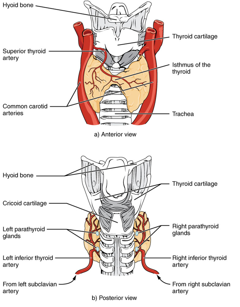
Hyoid bone The hyoid bone sits above the thyroid gland, offering a stable anchor for the tongue and larynx. It plays a supportive role in maintaining the neck’s structural integrity around the gland.
Thyroid cartilage The thyroid cartilage, known as the Adam’s apple, shields the larynx and supports vocal cord function. It frames the upper thyroid gland, providing a protective boundary.
Superior thyroid artery The superior thyroid artery delivers oxygenated blood to the thyroid’s upper region from the external carotid artery. This vessel is vital for sustaining the gland’s metabolic demands.
Common carotid arteries The common carotid arteries run alongside the thyroid, supplying blood to the head and neck. Their proximity ensures a robust circulation to support thyroid activity.
Isthmus of the thyroid The isthmus of the thyroid connects the gland’s two lobes across the trachea, forming a bridge-like structure. This connection allows the thyroid to encase the windpipe effectively.
Trachea The trachea, or windpipe, lies centrally beneath the thyroid, channeling air to the lungs. Its position enables the thyroid to influence respiratory and hormonal interactions.
Cricoid cartilage The cricoid cartilage forms the base of the larynx, sitting below the thyroid cartilage. It provides additional support and stability to the thyroid’s lower structure.
Left parathyroid glands The left parathyroid glands, located on the thyroid’s posterior surface, regulate calcium levels via parathyroid hormone. Their small size belies their critical role in mineral balance.
Right parathyroid glands The right parathyroid glands mirror the left, secreting hormones to maintain blood calcium. Their bilateral placement ensures symmetrical endocrine control.
Left inferior thyroid artery The left inferior thyroid artery, branching from the subclavian artery, nourishes the lower left lobe. It complements the superior artery for comprehensive blood supply.
Right inferior thyroid artery The right inferior thyroid artery supplies the lower right lobe, originating from the subclavian artery. This artery ensures balanced perfusion across the gland.
From left subclavian artery The left subclavian artery feeds the left inferior thyroid artery, delivering blood from the heart. This connection highlights the thyroid’s extensive vascular network.
From right subclavian artery The right subclavian artery supports the right inferior thyroid artery, maintaining symmetry in blood flow. This dual source enhances the gland’s functional resilience.
Anatomical Overview of the Thyroid Gland
The thyroid gland’s strategic location in the neck is essential for its endocrine role. Its butterfly-like shape, formed by two lobes and the isthmus, wraps around the trachea for optimal hormone distribution.
- The hyoid bone and thyroid cartilage provide upper structural support, protecting the gland.
- The common carotid arteries and superior thyroid artery ensure a rich blood supply.
- The trachea’s central position allows the thyroid to influence metabolic and respiratory functions.
- The cricoid cartilage stabilizes the lower gland, enhancing its anatomical fit.
Anterior View: Key Structures and Relationships
The anterior view showcases the thyroid’s external landmarks and vascular connections. This perspective highlights its interaction with major neck structures.
- The thyroid cartilage and hyoid bone frame the gland’s upper boundary, offering protection.
- The superior thyroid artery is visible, branching to supply the upper lobes.
- The isthmus connects the lobes, crossing the trachea for functional unity.
- Common carotid arteries run parallel, supporting overall neck circulation.
Posterior View: Deeper Anatomical Insights
The posterior view reveals the thyroid’s backside and associated glands. This angle provides insight into its blood supply and endocrine neighbors.
- The cricoid cartilage supports the lower thyroid, anchoring it to the larynx.
- Left and right parathyroid glands regulate calcium, nestled against the thyroid.
- The left and right inferior thyroid arteries originate from subclavian arteries.
- The bilateral blood supply from subclavian arteries ensures robust oxygenation.
Physiological Role and Blood Supply
The thyroid gland produces hormones like T3 and T4, which regulate metabolism and growth. Its extensive vascular network supports this critical function.
- The superior and inferior thyroid arteries provide dual blood sources, enhancing hormone synthesis.
- T3 (triiodothyronine) increases cellular metabolism, while T4 (thyroxine) acts as a prohormone.
- The parathyroid glands’ proximity aids in calcium regulation, complementing thyroid activity.
- Adequate blood flow prevents hypoxia, ensuring consistent hormone release.
Clinical Relevance and Imaging Techniques
Understanding thyroid anatomy assists in diagnosing and treating related conditions. Imaging and surgical planning rely on these anatomical details.
- The parathyroid glands’ location requires precision during thyroid surgery to avoid damage.
- Ultrasound assesses blood flow from carotid and subclavian arteries for vascular health.
- The isthmus’s position is key in evaluating goiter or nodule formation.
- Structural imaging helps detect abnormalities like thyroid enlargement or masses.
The thyroid gland’s intricate anatomy, supported by its vascular and structural relationships, underscores its pivotal role in endocrine health. Exploring its anterior and posterior views offers valuable insights into its function, aiding in both clinical practice and educational understanding of this essential organ.

