The carotid artery system serves as the primary conduit for oxygenated blood traveling from the heart to the brain and head. This essential vascular network ensures that the most metabolically demanding organs in the body receive a constant supply of nutrients to maintain consciousness and vital functions. This guide explores the anatomy of the common, internal, and external carotid arteries and their critical role in neurovascular health.
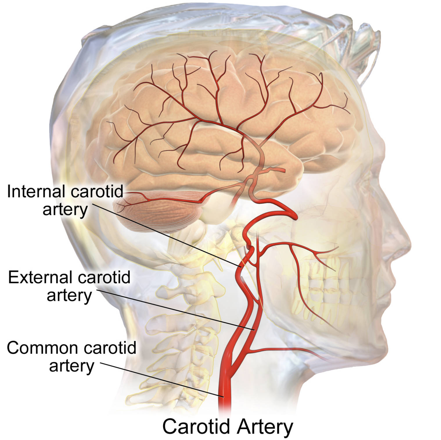
Internal carotid artery: This major branch travels upward into the cranium to provide the primary blood supply for the brain and eyes. It is a critical component of the Circle of Willis, ensuring collateral circulation to vital neurological tissues in the event of minor obstructions elsewhere.
External carotid artery: Unlike its internal counterpart, this artery remains outside the skull and branches into several smaller vessels to nourish the face, scalp, and neck organs. It plays a significant role in the vascularization of structures like the thyroid gland, facial muscles, and the tongue.
Common carotid artery: Originating from the aortic arch or brachiocephalic trunk, this vessel ascends the neck as a single trunk before splitting into its two main branches. It is easily palpable as a pulse in the neck and serves as the foundational pathway for the upper arterial system.
The carotid arteries are paired structures located on either side of the neck, acting as the highway for cerebrovascular blood flow. Without a steady supply of oxygen and nutrients from these vessels, brain function would cease within minutes. The bifurcation point, where the common artery splits into the internal and external branches, is a particularly important anatomical landmark, housing the carotid sinus and carotid body.
These structures act as sensitive pressure and chemical sensors. The carotid sinus contains baroreceptors that monitor blood pressure, while the carotid body contains chemoreceptors that sense oxygen and carbon dioxide levels in the blood. This allows the body to make rapid cardiovascular adjustments to maintain homeostasis and proper respiratory rates.
The anatomical path of these vessels is crucial for several clinical reasons:
- It facilitates the high-pressure delivery of blood needed for complex cognitive functions.
- The branching pattern allows for specialized delivery to either deep internal brain structures or superficial facial features.
- The vessel’s accessible location in the neck makes it a primary site for clinical pulse assessments and vascular surgeries like endarterectomy.
Understanding this system is not just about anatomy; it is about recognizing the risk factors for conditions like carotid artery disease. This occurs when plaque builds up in the arterial walls, leading to stenosis or narrowing of the vessel lumen. If a piece of this plaque breaks off or a blood clot forms at the site, it can travel to the brain, causing an ischemic stroke.
The Physiological Pathway of the Carotid System
The physiology of the carotid system is designed for both efficiency and protection. The common carotid artery on the left side arises directly from the aorta, while the right common carotid begins at the brachiocephalic trunk. As these vessels ascend the neck, they are encased and protected by the carotid sheath, a layer of connective tissue that also houses the internal jugular vein and the vagus nerve.
When blood reaches the bifurcation, usually at the level of the upper border of the thyroid cartilage, the internal carotid artery takes a more posterior path. It enters the skull via the carotid canal of the temporal bone. Once inside, it branches further into the anterior and middle cerebral arteries. These vessels are responsible for supplying the vast majority of the cerebral hemispheres. If the internal carotid becomes narrowed, the brain relies on its unique vascular ring at the base of the skull to reroute blood from the vertebral arteries or the opposite carotid artery.
Clinical Significance of the Carotid Bifurcation
The external carotid artery, conversely, splits into eight main branches, including the superior thyroid, lingual, facial, and maxillary arteries. These are essential for everything from speech and swallowing to maintaining the health of the skin on the scalp. Because the face has such a rich blood supply, injuries to this area often bleed profusely, which is a testament to the high volume of blood carried by the external carotid system.
The carotid bifurcation is the most common site for atherosclerosis in the neck. The turbulent blood flow at the “Y” junction of the artery can cause micro-injuries to the vessel lining, which facilitates the accumulation of cholesterol and inflammatory cells. Managing vascular health through a balanced diet, regular exercise, and tobacco cessation is vital for preventing the narrowing of these critical vessels and ensuring long-term brain health.
The carotid artery system is a masterpiece of vascular engineering, balancing the immense pressure of the heart’s output with the delicate needs of the human brain. By maintaining a clear understanding of its anatomy—from the common trunk to the specialized internal and external branches—we can better appreciate the complexities of the human body and the importance of vascular health. Protecting these vessels through a healthy lifestyle is one of the most effective ways to ensure long-term neurological and systemic well-being.

