The human heart, a vital organ responsible for circulating blood throughout the body, resides within the protective confines of the thoracic cavity. This intricate image provides a clear anatomical overview of the heart’s position relative to surrounding structures, including the lungs, major blood vessels, and diaphragm. Understanding this spatial arrangement is crucial for comprehending cardiovascular function and identifying potential health concerns.
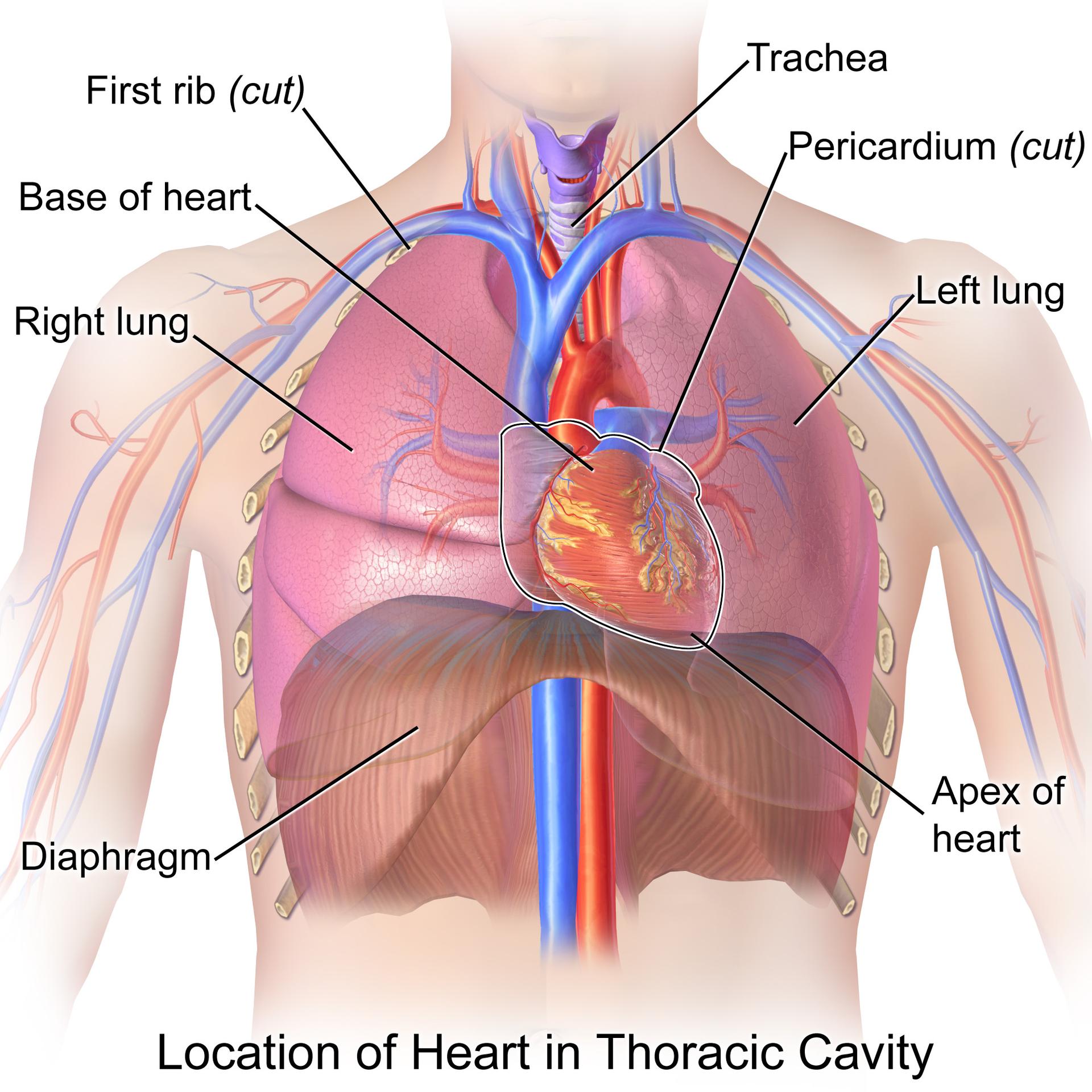
First rib (cut): The first rib is the uppermost bone of the rib cage, playing a crucial role in protecting the apex of the lungs and the major vessels of the neck. In this diagram, it is depicted as “cut” to allow for a clearer view of the underlying structures.
Trachea: The trachea, commonly known as the windpipe, is a cartilaginous tube that extends from the larynx and branches into the bronchi, facilitating the passage of air to and from the lungs. Its anterior position to the esophagus highlights its direct role in the respiratory system.
Pericardium (cut): The pericardium is a double-layered sac that encloses the heart, providing protection and anchoring it within the mediastinum. The “cut” view in the image allows us to observe the heart’s outer surface, demonstrating how this protective sac is typically situated.
Base of heart: The base of the heart refers to its superior aspect, where the great blood vessels, such as the aorta and pulmonary arteries and veins, enter and exit the organ. This region is broader and less pointed compared to the apex.
Right lung: The right lung is one of the two main respiratory organs, responsible for gas exchange. It is typically larger than the left lung and is divided into three lobes.
Left lung: The left lung, slightly smaller than the right to accommodate the heart, is also a primary organ of respiration, divided into two lobes. Its position demonstrates the heart’s slight leftward orientation within the chest.
Diaphragm: The diaphragm is a large, dome-shaped muscle located at the base of the thoracic cavity, crucial for respiration. Its contraction and relaxation facilitate the expansion and compression of the lungs during breathing.
Apex of heart: The apex of the heart is the pointed, inferior portion of the heart that typically points towards the left hip. It is formed by the left ventricle and rests on the diaphragm, producing the apical impulse that can be felt on the chest wall.
The heart, a remarkable muscular pump, is strategically positioned within the thoracic cavity, an area often referred to as the chest. This vital organ is nestled between the lungs, slightly to the left of the midline, and superior to the diaphragm. Its precise location is within the mediastinum, the central compartment of the thoracic cavity. This protective environment ensures the heart’s functionality while shielding it from external impact. The anatomical arrangement, as illustrated, underscores the close relationship between the cardiovascular and respiratory systems, with major vessels and airways in immediate proximity to the heart.
Understanding the specific placement of the heart is not merely an academic exercise; it holds significant implications for medical diagnostics and interventions. For instance, the apex of the heart provides a key landmark for auscultation, allowing clinicians to listen to heart sounds. Similarly, knowledge of the pericardium’s protective role is essential in understanding conditions like pericarditis. The heart’s intricate connection to the surrounding structures, including the major arteries and veins emanating from its base, highlights its central role in systemic circulation.
- The heart is about the size of a clenched fist.
- It pumps approximately 5 liters of blood per minute.
- The pericardium reduces friction during heart contractions.
- The diaphragm is essential for normal breathing mechanics.
This detailed anatomical perspective serves as a foundational element for both medical professionals and individuals seeking to understand the complexities of the human body. The consistent pumping action of the heart, facilitated by its protected location and intricate connections, is fundamental to life. Recognizing these relationships can aid in the early detection and management of various cardiovascular and respiratory conditions, emphasizing the importance of a holistic view of the human body’s interconnected systems.
The precise positioning of the heart within the thoracic cavity, as clearly depicted in this image, is a testament to the efficient design of the human body. Each surrounding structure, from the protective ribs and pericardium to the vital trachea and lungs, plays a crucial role in supporting the heart’s function and overall physiological balance. This anatomical understanding is not only foundational for medical studies but also empowers individuals with a greater appreciation for the intricate mechanisms that sustain life.

