The thymus serves as a critical training ground where immature T cells, known as thymocytes, undergo a transformative journey to become functional components of the adaptive immune system. Located in the upper chest, this organ facilitates a series of developmental stages that ensure thymocytes develop both functionality and self-tolerance before being released into circulation. This detailed illustration captures the intricate process of T cell maturation, offering a window into the mechanisms that shape immune competence.
Labeled Components of T Cell Differentiation
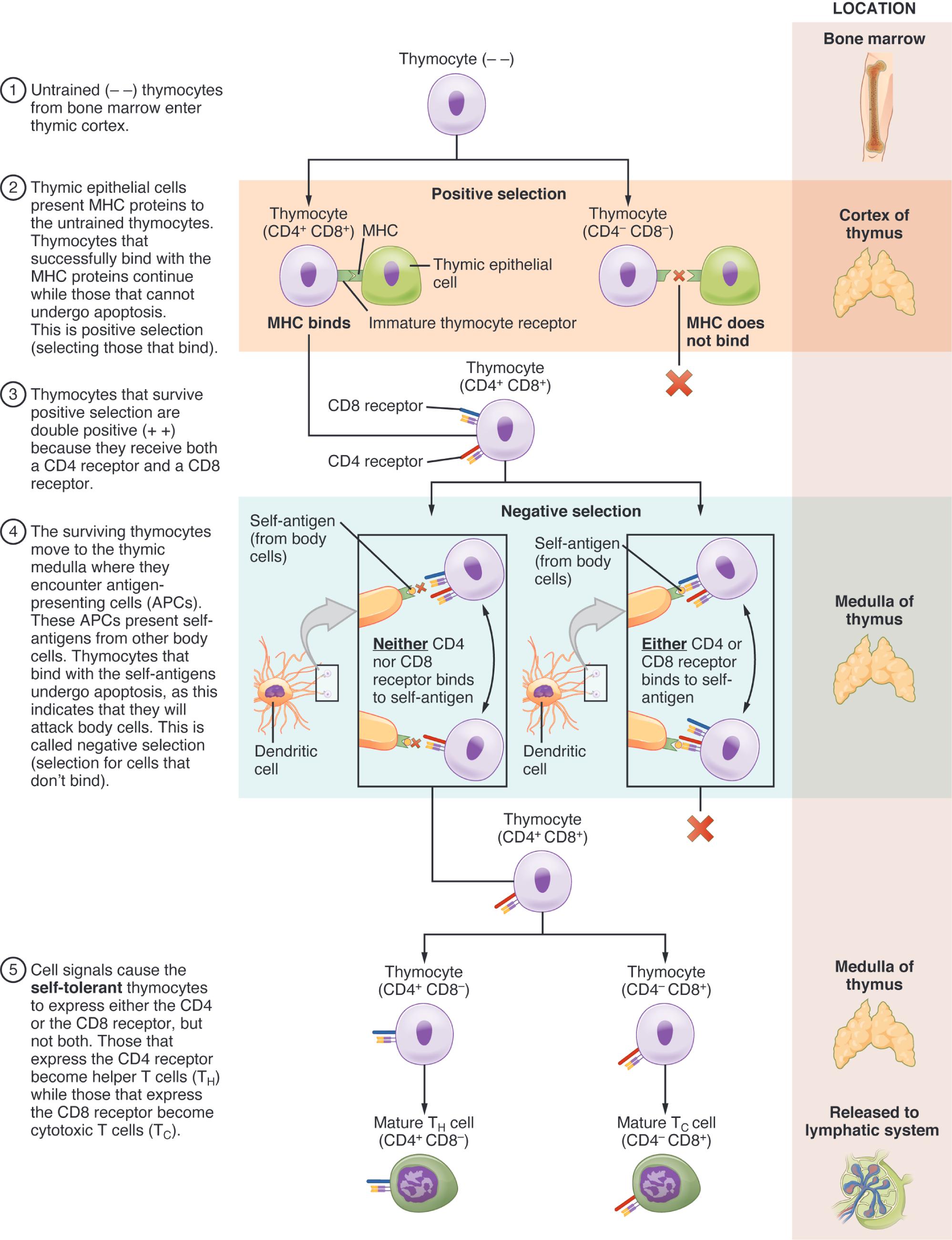
Thymocyte (immature): These precursor cells enter the thymus from the bone marrow, lacking specificity or tolerance. They undergo proliferation and selection to mature into functional T cells.
Thymocyte (double-negative): Lacking CD4 and CD8 co-receptors, these early thymocytes begin T cell receptor (TCR) gene rearrangement. They represent the initial stage of T cell development in the cortex.
Thymocyte (double-positive): Expressing both CD4 and CD8, these thymocytes undergo positive and negative selection in the cortex. They test their TCRs for functionality and self-tolerance.
Thymocyte (single-positive CD4+): After selection, these cells express only CD4, becoming helper T cells. They exit the thymus to coordinate immune responses against extracellular pathogens.
Thymocyte (single-positive CD8+): These cells, expressing only CD8 post-selection, mature into cytotoxic T cells. They leave the thymus to target and destroy infected or abnormal cells.
Cortex: The outer region of the thymus, rich in epithelial cells, supports thymocyte proliferation and early selection. It is where double-negative and double-positive stages occur.
Medulla: The inner region contains mature thymocytes and Hassall’s corpuscles, facilitating final tolerance checks. It is the site where single-positive cells are prepared for release.
Epithelial cell: These thymic cells provide a supportive microenvironment for thymocyte development. They express self-antigens to test T cell tolerance during selection.
Dendritic cell: Present in the medulla, these cells present self-antigens to thymocytes. They play a key role in negative selection to eliminate self-reactive T cells.
Macrophage: These cells phagocytose apoptotic thymocytes that fail selection. They maintain a clean thymic environment, supporting healthy T cell maturation.
Positive selection: This process ensures thymocytes with functional TCRs that recognize MHC molecules survive. It occurs in the cortex, shaping the T cell repertoire.
Negative selection: Taking place in the medulla, this step eliminates thymocytes with strong self-reactivity. It prevents autoimmunity by removing potentially harmful cells.
Apoptosis: This programmed cell death removes thymocytes that fail selection criteria. It is a natural pruning process to refine the T cell population.
Mature T cell: These fully developed cells, either CD4+ or CD8+, exit the thymus to join the peripheral immune system. They are ready to respond to specific antigens.
Anatomical Context of T Cell Differentiation
The thymus provides a structured environment for T cell maturation, with distinct regions driving development.
- Thymocytes enter as immature cells, initiating their journey in the cortex.
- The cortex hosts double-negative and double-positive stages, supported by epithelial cells.
- The medulla refines single-positive thymocytes, aided by dendritic cells and macrophages.
- Positive selection shapes functional TCRs, while negative selection ensures tolerance.
- Apoptosis clears unfit cells, maintaining immune balance.
- Mature T cells exit via blood vessels to patrol the body.
This illustration highlights the thymus’s role as a maturation hub.
Physiological Role in Immune Development
T cell differentiation equips the immune system with a diverse and tolerant T cell pool.
- Immature thymocytes proliferate, generating a large pool for selection.
- Double-negative thymocytes rearrange TCR genes, establishing diversity.
- Double-positive thymocytes undergo selection, ensuring MHC recognition.
- Single-positive CD4+ cells develop into helpers, activating other immune cells.
- Single-positive CD8+ cells become cytotoxic, targeting infected cells.
- Epithelial and dendritic cells guide the process, promoting tolerance.
This maturation ensures a balanced immune response.
Mechanisms of Selection Processes
The thymus employs rigorous checks to produce effective T cells.
- Positive selection tests TCR-MHC binding, retaining viable thymocytes.
- Negative selection uses self-antigens to eliminate autoreactive cells.
- Epithelial cells in the cortex support positive selection with MHC expression.
- Dendritic cells in the medulla present self-antigens for negative selection.
- Macrophages remove apoptotic cells, preventing inflammation.
- This dual selection refines the T cell repertoire for functionality.
These mechanisms are critical for immune competence.
Clinical Relevance of T Cell Differentiation
Understanding thymic processes aids in managing immune disorders.
- Thymic aplasia, as in DiGeorge syndrome, leads to T cell deficiency.
- Abnormal positive selection can cause immune hyporesponsiveness.
- Negative selection failures contribute to autoimmune diseases like lupus.
- Cortical or medullary damage may impair thymocyte maturation.
- Macrophage dysfunction can lead to thymic inflammation.
- Thymic function decline with age affects T cell production.
This knowledge guides diagnostic and therapeutic approaches.
Developmental and Adaptive Features
T cell differentiation adapts to the body’s evolving immune needs.
- Thymocytes mature through stages, peaking in childhood for diversity.
- The cortex and medulla refine selection as the thymus grows.
- Epithelial cells adapt antigen presentation to match self-tissues.
- Dendritic cells evolve to enhance tolerance with age.
- Apoptosis rates adjust to maintain T cell quality.
- Mature T cells develop memory potential for future responses.
This adaptability ensures lifelong immune protection.
The thymus’s role in T cell differentiation, as depicted in this illustration, is a fascinating process that transforms immature thymocytes into a diverse, tolerant T cell population. By guiding these cells through selection and maturation, the thymus lays the foundation for a robust adaptive immune response, making it an essential focus for exploring immune health and development.

