Understanding the Female Reproductive System: Anatomy Guide

Date:

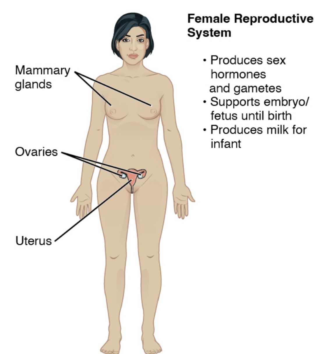
The female reproductive system is a sophisticated network of organs designed for reproduction, hormonal regulation, and menstrual cycles, as illustrated in the provided image. This article offers a detailed exploration of the anatomical structures depicted, shedding light on their functions and interconnections. By examining this system, one can gain a deeper appreciation of its critical role in fertility and overall health.
Understanding the Female Reproductive System: Anatomy Guide

Ovaries: These almond-shaped organs produce eggs (ova) and hormones like estrogen and progesterone. They play a central role in ovulation and maintaining the menstrual cycle.

Fallopian tubes: These tubes extend from the ovaries to the uterus, serving as pathways for eggs to travel after ovulation. They are also the site where fertilization typically occurs if sperm is present.

- Advertisement -

Recommended Study Resource

Gray's Anatomy: The Anatomical Basis of Clinical Practice

Enhance your anatomical knowledge with Gray's Anatomy: The Anatomical Basis of Clinical Practice. This authoritative text offers in-depth insights and illustrations, perfect for medical students and practitioners aiming for clinical excellence.

Shop Now on Amazon

At AnatomyNote.com, we offer free resources on anatomy, pathology, and pediatric medicine for medical students and professionals. Purchasing through our Amazon links, like Gray's Anatomy, supports our server costs and content creation at no additional cost to you.

Disclosure: As an Amazon Associate, we earn a commission from qualifying purchases.

Disclosure: As an Amazon Associate, we earn a commission from qualifying purchases at no extra cost to you.

Uterus: This pear-shaped organ houses and nourishes a developing fetus during pregnancy. Its muscular walls expand significantly to accommodate fetal growth.

Cervix: The lower part of the uterus, this narrow passage connects to the vagina and produces mucus to protect the reproductive tract. It dilates during childbirth to allow the baby to pass.

Vagina: This muscular canal serves as the birth canal and the exit for menstrual flow. It also facilitates sexual intercourse and is lined with mucus membranes for protection.

Endometrium: The inner lining of the uterus, this tissue thickens each month in preparation for pregnancy. It sheds during menstruation if no fertilization occurs.

- Advertisement -

Anatomy Flash Cards

Master anatomy with detailed, exam-ready flash cards.

Buy on Amazon

AnatomyNote.com offers free anatomy and pathology resources. Your purchase of Anatomy Flash Cards supports our site at no extra cost.

As an Amazon Associate, we earn from qualifying purchases.

Anatomical Structure of the Female Reproductive System

The female reproductive system’s anatomy is intricately designed to support reproduction. Each component contributes uniquely to this process.

  • The ovaries contain thousands of follicles, each potentially releasing an egg.
  • The fallopian tubes are lined with cilia to help move the egg toward the uterus.
  • The uterus has three layers: the endometrium, myometrium, and perimetrium.
  • The cervix contains a small opening, the os, which varies in size during the cycle.
  • The vagina is supported by pelvic floor muscles for structural integrity.

Physiological Functions and Hormonal Regulation

The physiological roles of the female reproductive system are driven by hormonal changes. This dynamic process supports reproduction and monthly cycles.

  • The ovaries release estrogen and progesterone, regulating the menstrual cycle.
  • The fallopian tubes provide an environment for fertilization by the sperm.
  • The uterus contracts during labor, aided by progesterone withdrawal.
  • The cervix produces fertile mucus during ovulation to aid sperm passage.
  • The endometrium regenerates monthly under hormonal influence.

Clinical Relevance and Health Considerations

While the image focuses on anatomy, understanding the female reproductive system aids in recognizing potential health issues. Regular screening can prevent complications.

  • The ovaries are prone to cysts or tumors, requiring ultrasound monitoring.
  • Blockages in the fallopian tubes can lead to infertility, treatable with surgery.
  • The uterus may develop fibroids, affecting menstrual flow or pregnancy.
  • The cervix is screened for cervical cancer via Pap smears.
  • Changes in the endometrium can indicate conditions like endometriosis.

The female reproductive system, as depicted, highlights the coordinated functions of the ovaries, fallopian tubes, uterus, cervix, vagina, and endometrium in supporting reproduction. This anatomical guide provides a foundation for exploring its physiological roles and clinical significance. By studying these structures, one can better understand their importance in maintaining health and addressing reproductive challenges effectively.

We'd be thrilled to have your support!

Your generous contribution through a coffee keeps our passion alive.

Image source:
  • Authors: J. Gordon Betts, Kelly A. Young, James A. Wise, Eddie Johnson, Brandon Poe, Dean H. Kruse, Oksana Korol, Jody E. Johnson, Mark Womble, Peter DeSaix

LEAVE A REPLY

Please enter your comment!
Please enter your name here

Share post:

Popular

spot_imgspot_img

Subscribe

More like this
Related

Female Reproductive System: Detailed Anatomical Structure and Function

The female reproductive system comprises a complex network of internal and external organs designed for reproduction, hormone production, and sexual function. This detailed anatomical guide presents a comprehensive overview of all reproductive structures, from the fallopian tubes to the external genitalia, with particular emphasis on their anatomical relationships and physiological functions. Understanding these structures is crucial for medical professionals in diagnosing and treating reproductive conditions.

Human Female Internal Genitalia and Penis Max Penetration: A Comprehensive Anatomical Guide

The female reproductive system comprises a complex network of organs and structures designed for reproduction, hormone production, and sexual function. Each component demonstrates specific anatomical features and physiological roles essential for reproductive health. Understanding these structures in detail is crucial for medical professionals in diagnosing and treating various gynecological conditions.

Human Egg Cell Anatomy: Detailed Structure of the Ovum Explained

The human egg cell, or ovum, is a critical component of reproduction, serving as the female gamete in the fertilization process. This article explores a labeled diagram of the ovum, detailing its anatomical structure and the roles of its components in human reproduction. Ideal for medical students, healthcare professionals, and those interested in reproductive biology, this guide offers a comprehensive look at the ovum's anatomy and its significance in conception.

Female Reproductive Organs Diagram with labels

The female reproductive system is a complex network of organs and structures designed for reproduction, hormone production, and menstruation. This anatomical diagram illustrates the key components of the internal reproductive organs, including the uterus, ovaries, fallopian tubes, and associated structures, providing essential insights into female reproductive health and function.