Explore the crucial anatomical relationship between the human heart and the protective rib cage, as depicted in this detailed illustration. This spatial understanding is vital for both medical professionals and those interested in human anatomy, offering insights into how the body safeguards one of its most critical organs. Grasping the heart’s precise location relative to the ribs is fundamental for diagnostics and understanding physical protection.
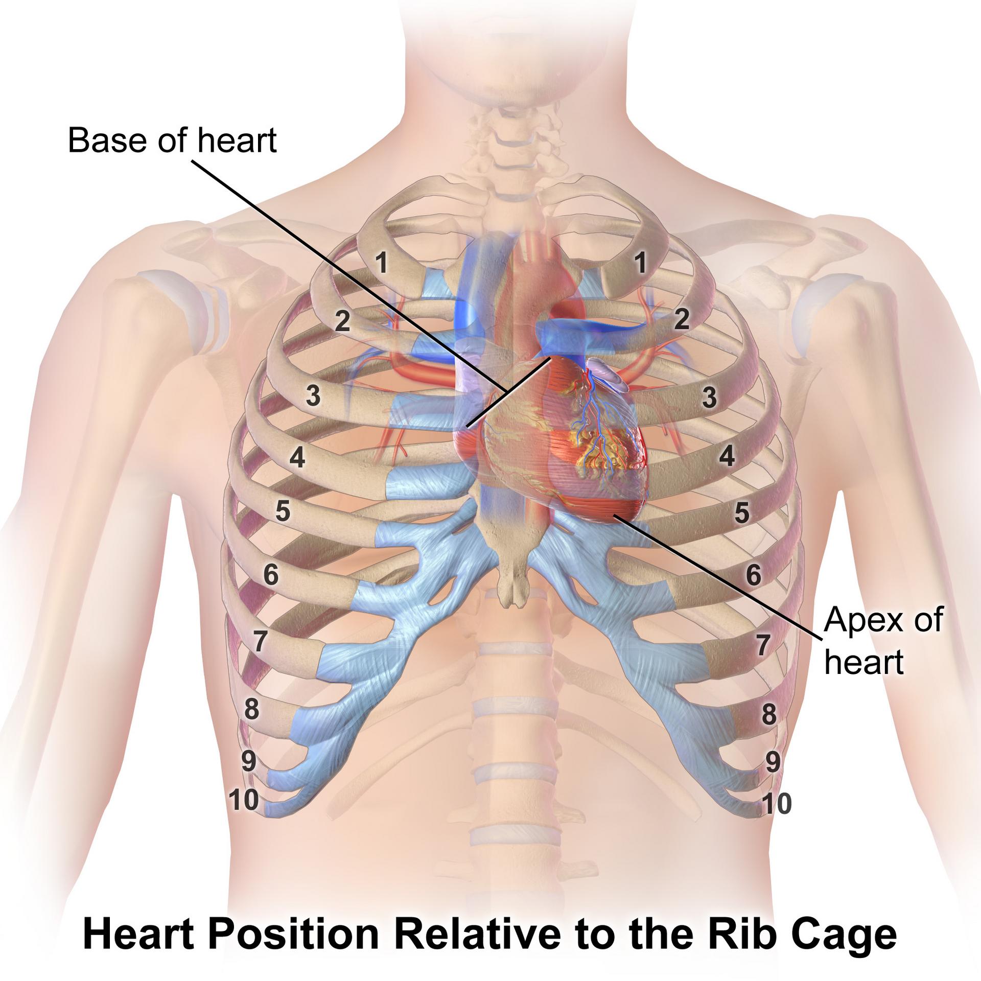
Base of heart: The base of the heart refers to its broader, superior aspect, from which the great vessels, such as the aorta and pulmonary arteries, emerge. It is typically situated at the level of the second or third costal cartilages, largely obscured by the sternum and ribs.
Apex of heart: The apex of the heart is the pointed, inferior portion of the organ, primarily formed by the left ventricle. It typically points downwards, forwards, and to the left, often lying in the fifth intercostal space, about 8-9 cm from the midsternal line.
Ribs (1-10): The ribs are curved bones that form the rib cage, playing a vital role in protecting internal organs such as the heart and lungs. Numbered 1 through 12, the illustration highlights the first ten pairs, demonstrating how they encase and shield the thoracic cavity.
The human heart, a ceaseless pump essential for life, is remarkably well-protected within the thoracic cavity by the rib cage. This bony structure acts as a natural shield, safeguarding the heart from external trauma and impacts. The illustration clearly shows how the ribs, sternum, and vertebral column form a robust enclosure around the heart, providing a stable environment for its continuous operation. Understanding this anatomical relationship is crucial for interpreting medical imaging, performing physical examinations, and comprehending the biomechanics of chest injuries.
The heart’s position is not entirely symmetrical; it lies slightly to the left of the midline, with its apex pointing towards the left hip. This orientation explains why heart sounds are often best heard on the left side of the chest. The base of the heart, where major blood vessels connect, is superiorly located, primarily behind the sternum and upper ribs. This strategic placement within the rib cage ensures that while it is protected, it also remains central enough to effectively distribute blood to both sides of the body.
The precise location of the heart’s apex is a critical clinical landmark, often palpable as the point of maximal impulse (PMI). This landmark, usually found in the fifth intercostal space at the midclavicular line, provides valuable diagnostic information regarding heart size and function. Any deviation in the PMI can indicate conditions such as cardiomegaly (enlarged heart). Therefore, the relationship between the heart’s structures and the surrounding bony framework is not just descriptive but has significant diagnostic and prognostic implications in cardiology.
- The rib cage consists of 12 pairs of ribs.
- The sternum and vertebral column complete the protective cage.
- The heart is roughly the size of a clenched fist.
- The rib cage allows for expansion and contraction during respiration.
This detailed visual of the heart’s position relative to the rib cage underscores the intricate design of the human body, where protection and function are meticulously integrated. The robust bony architecture serves to safeguard the delicate cardiac muscle, ensuring its uninterrupted performance. Such anatomical insights are fundamental for healthcare professionals, aiding in accurate diagnoses and treatments, and for anyone seeking to understand the remarkable protective mechanisms of human physiology.
The detailed depiction of the heart’s position within the rib cage highlights the vital protective role of the skeletal system for our most crucial organs. This anatomical understanding is essential for grasping how the heart is shielded from external forces while maintaining its central function in circulating blood. A clear comprehension of this relationship is fundamental for medical practitioners and for anyone interested in the intricate design of the human body.

