Explore the elegant simplicity and remarkable efficiency of the two-chambered heart found in fish, a cardiovascular design perfectly adapted for aquatic environments. This article delves into the unique single-circuit circulatory system that ensures continuous blood flow through the gills for oxygenation and then to the rest of the body. Understand how this fundamental cardiac structure supports the diverse physiological demands of piscine life.
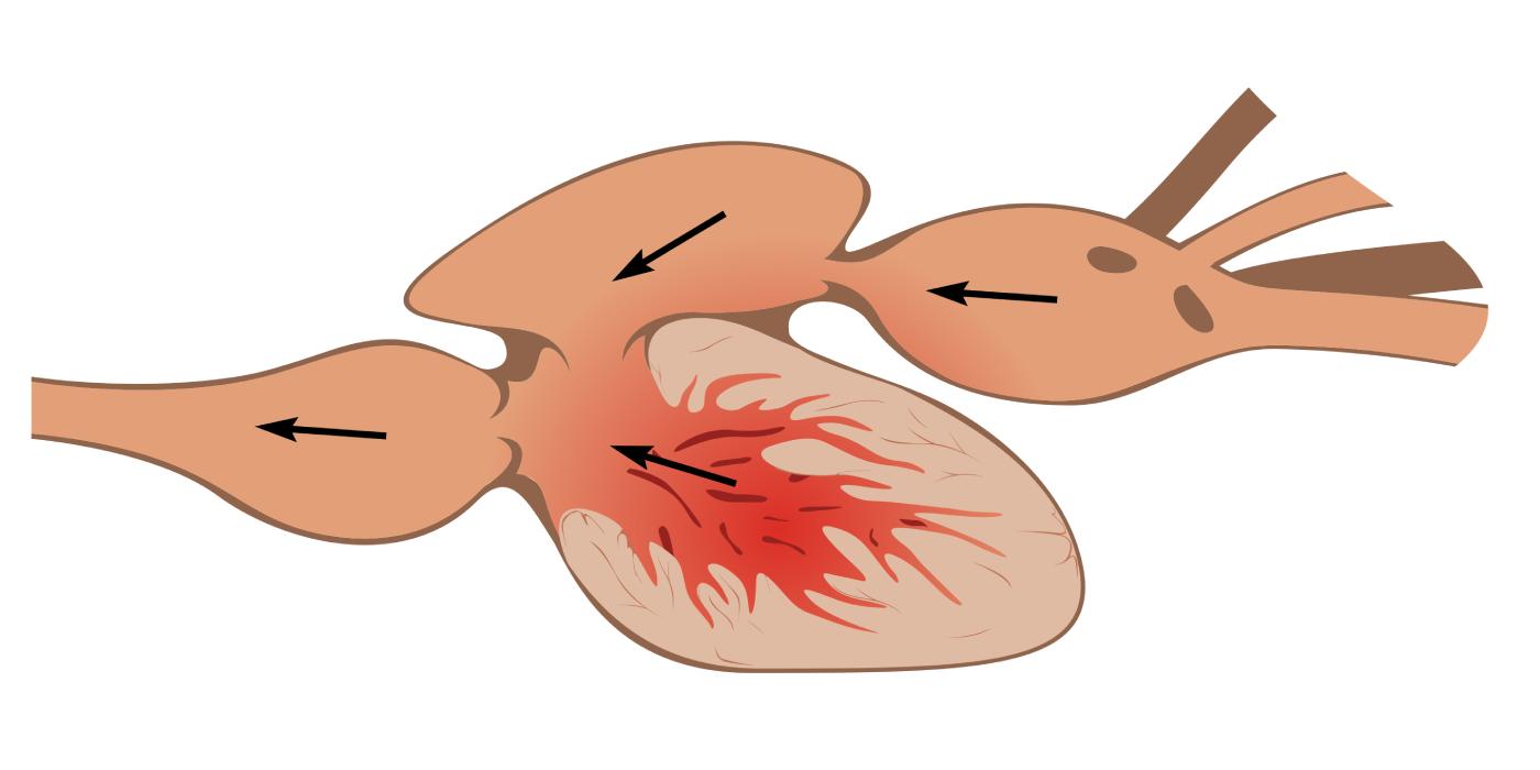
The cardiovascular system of fish is uniquely adapted to their aquatic existence, featuring a distinctive two-chambered heart that drives a single-circuit circulatory pathway. Unlike the double-circuit systems found in terrestrial vertebrates, where blood is pumped separately to the lungs and the body, a fish’s heart propels blood first through the gills for oxygenation, and then directly to the rest of the body. This efficient design ensures that every pump of the heart contributes directly to both respiration and systemic circulation, optimizing oxygen delivery in a water-bound environment.
The two-chambered heart consists of a single atrium and a single ventricle, arranged in series. Deoxygenated blood from the body enters the atrium, which then pumps it into the muscular ventricle. The ventricle, being the primary pumping chamber, generates the force necessary to push the blood forward. From the ventricle, the blood flows into a series of specialized structures that guide it towards the gills. This unidirectional flow is a hallmark of the fish circulatory system, ensuring a continuous and orderly progression of blood throughout the body.
Upon leaving the heart, the blood travels to the gills, where it undergoes gas exchange, releasing carbon dioxide and absorbing oxygen from the water. Once oxygenated, the blood continues its journey directly to the systemic capillaries throughout the body, delivering oxygen and nutrients to tissues and collecting metabolic waste products. This process, where the gills act as both the respiratory and initial systemic capillary beds, results in a drop in blood pressure after leaving the gills. Despite this, the system is highly effective for fish, supporting their metabolic needs and diverse active lifestyles in aquatic habitats.
Key components of the fish circulatory system include:
- Two-chambered heart: Single atrium and single ventricle.
- Single-circuit circulation: Blood flows in one continuous loop.
- Gills: Site of gas exchange.
- Unidirectional blood flow: Ensures efficient transport.
- Systemic capillaries: Deliver oxygen to body tissues.
In conclusion, the two-chambered heart of fish represents a highly specialized and efficient cardiovascular design, perfectly tailored for life in water. Its single-circuit circulatory system, moving deoxygenated blood from the body to the atrium and ventricle, then to the gills for oxygenation, and finally to the rest of the body, underscores a fundamental principle of adaptation. This elegant anatomical solution allows fish to thrive in diverse aquatic environments, demonstrating how evolutionary pressures shape even the most vital physiological systems to meet specific environmental demands.

