The cerebellum stands as a critical brain region dedicated to motor coordination, balance, and cognitive functions, positioned posterior to the brain stem. This anatomical illustration depicts key structures including the cerebellum, pons, inferior olive, and deep cerebellar white matter (arbor vitae), illustrating input and output pathways essential for precise movement control. Accompanied by an imaging view, this representation offers valuable insights into cerebellar organization and its integration with surrounding neural elements.
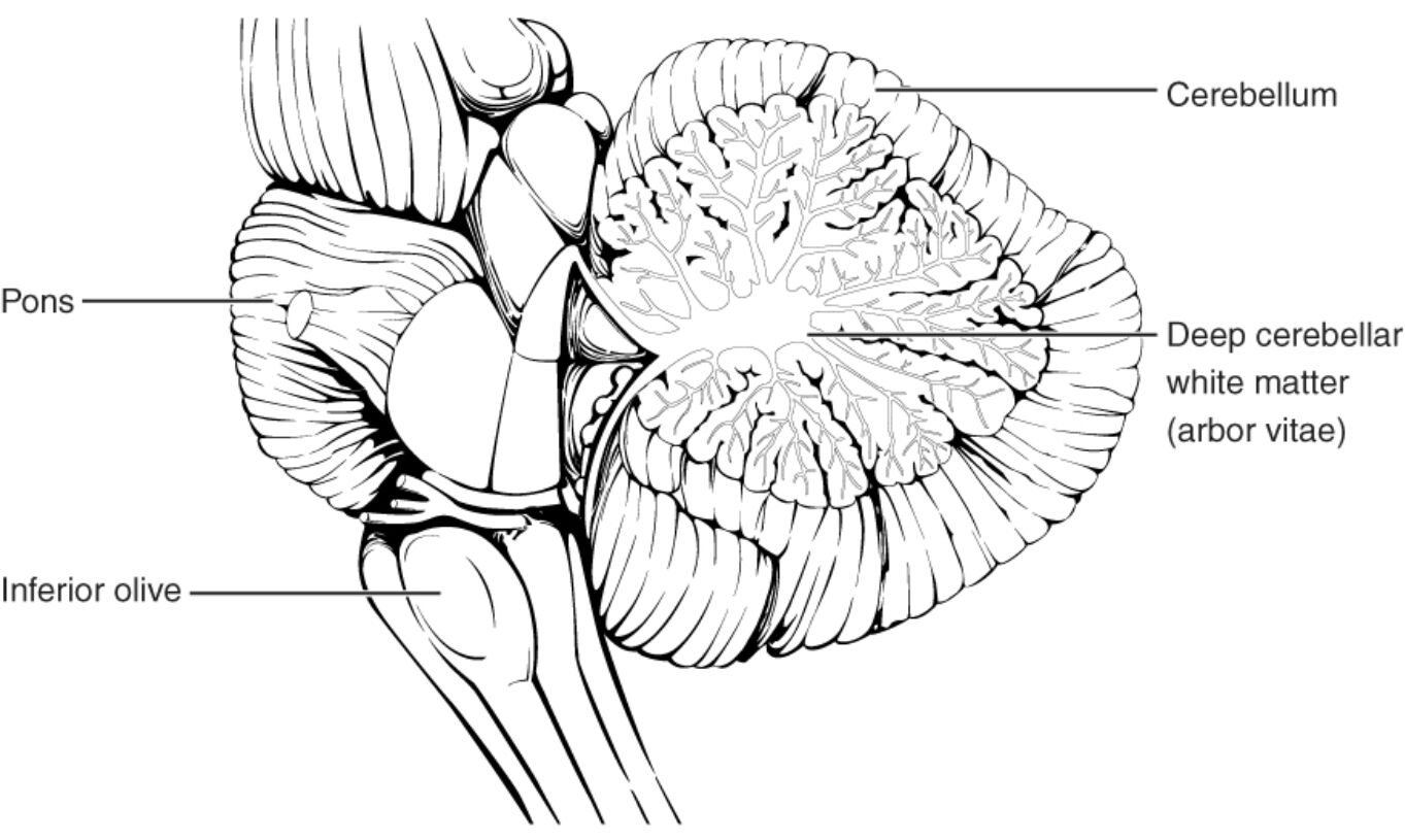
Cerebellum
The cerebellum, located behind the brain stem, refines voluntary movements, maintains equilibrium, and supports non-motor tasks such as language and attention. Its highly folded surface, or folia, maximizes cortical area for processing vast sensory inputs from the spinal cord and cerebral cortex.
Pons
The pons, a bulbous structure in the brain stem, facilitates communication between the cerebral cortex and cerebellum via large white matter tracts, enabling descending inputs for motor planning. It also houses nuclei for several cranial nerves, contributing to facial movements, hearing, and respiratory control.
Inferior olive
The inferior olive, a medullary nucleus, supplies climbing fibers to the cerebellum, crucial for error detection and motor learning through strong synaptic connections with Purkinje cells. These fibers convey ascending sensory information from the periphery and spinal cord, aiding adaptive movement refinement.
Deep cerebellar white matter (arbor vitae)
The deep cerebellar white matter, known as arbor vitae due to its tree-like branching, consists of myelinated axons interconnecting cerebellar cortex with deep nuclei and peduncles. This structure ensures efficient signal transmission, supporting the cerebellum’s role in coordinating outputs to the midbrain and beyond.
Anatomical Overview of the Cerebellum
The cerebellum integrates sensory and motor information to produce smooth actions. Its posterior positioning on the brain stem optimizes connectivity for real-time adjustments.
- The cerebellum comprises two hemispheres and a central vermis, with the hemispheres handling limb movements and the vermis focusing on trunk posture.
- Divided into anterior, posterior, and flocculonodular lobes, each lobe processes distinct inputs for gait, precision, and vestibular functions.
- Cerebellar peduncles—superior, middle, and inferior—link it to the midbrain, pons, and medulla, respectively.
- Arterial supply from branches of the basilar artery nourishes its high metabolic demands.
Detailed Examination of the Pons
The pons acts as a relay hub in the brain stem, bridging higher and lower neural centers. Its white matter prominence reflects extensive fiber tracts.
- Transverse pontine fibers form the middle cerebellar peduncle, conveying cortical commands for planned movements.
- Longitudinal tracts include corticospinal fibers for voluntary motor control and medial lemniscus for sensory relay.
- Pontine nuclei project to granule cells in the cerebellar cortex, amplifying input signals.
- Venous drainage occurs via the petrosal sinuses, maintaining circulatory balance.
Structure and Function of the Inferior Olive
The inferior olive features a convoluted, olive-like shape in the ventral medulla. It specializes in providing instructive signals to the cerebellum.
- Olivocerebellar fibers decussate and ascend through the inferior peduncle, terminating as climbing fibers on Purkinje dendrites.
- Electrotonic coupling via gap junctions synchronizes olivary neuron firing, generating complex spike patterns for timing.
- Inputs from the spinal cord via spino-olivary tracts and from the red nucleus integrate proprioceptive and descending data.
- Glutamatergic transmission excites Purkinje cells, facilitating long-term depression for learning.
Insights into Deep Cerebellar White Matter
The arbor vitae branching pattern symbolizes life’s tree in anatomical terms. It forms the core framework for cerebellar connectivity.
- Afferent fibers enter via peduncles, branching to distribute signals across folia.
- Efferent axons from deep nuclei exit through the superior peduncle, targeting the thalamus and red nucleus.
- Myelin integrity, assessed in imaging, correlates with conduction velocity up to 100 m/s.
- Oligodendrocyte support maintains axonal health, crucial for signal fidelity.
Imaging Representation of the Cerebellum
The lower imaging view highlights the cerebellum in purple, showcasing its appearance in diagnostic scans. This modality reveals internal architecture non-invasively.
- MRI contrasts differentiate gray matter folia from white matter tracts, with T2-weighted images emphasizing fluid spaces.
- The highlighted region displays the arbor vitae as hyperintense branches, aiding volume measurements.
- Functional imaging tracks activation during tasks, linking structure to performance.
- Diffusion imaging maps fiber orientations, identifying connectivity patterns.
Physiological Pathways in the Cerebellum
Input and output circuits define cerebellar operations. Descending and ascending pathways converge for output modulation.
- Cortical inputs via the pons reach mossy fibers, exciting granule cells and parallel fibers.
- Spinal inputs through the inferior olive activate climbing fibers for error signals.
- Purkinje cells inhibit deep nuclei, shaping outputs to the midbrain and spinal cord.
- GABAergic interneurons regulate circuit excitability, preventing overload.
Conclusion
The cerebellum’s anatomical structure, as shown in this illustration and imaging, underscores its pivotal role in neural harmony. Through connections via the pons and inferior olive, and the intricate arbor vitae, it exemplifies precision in brain function. This detailed view not only illuminates foundational anatomy but also enhances appreciation for its contributions to movement and cognition.

