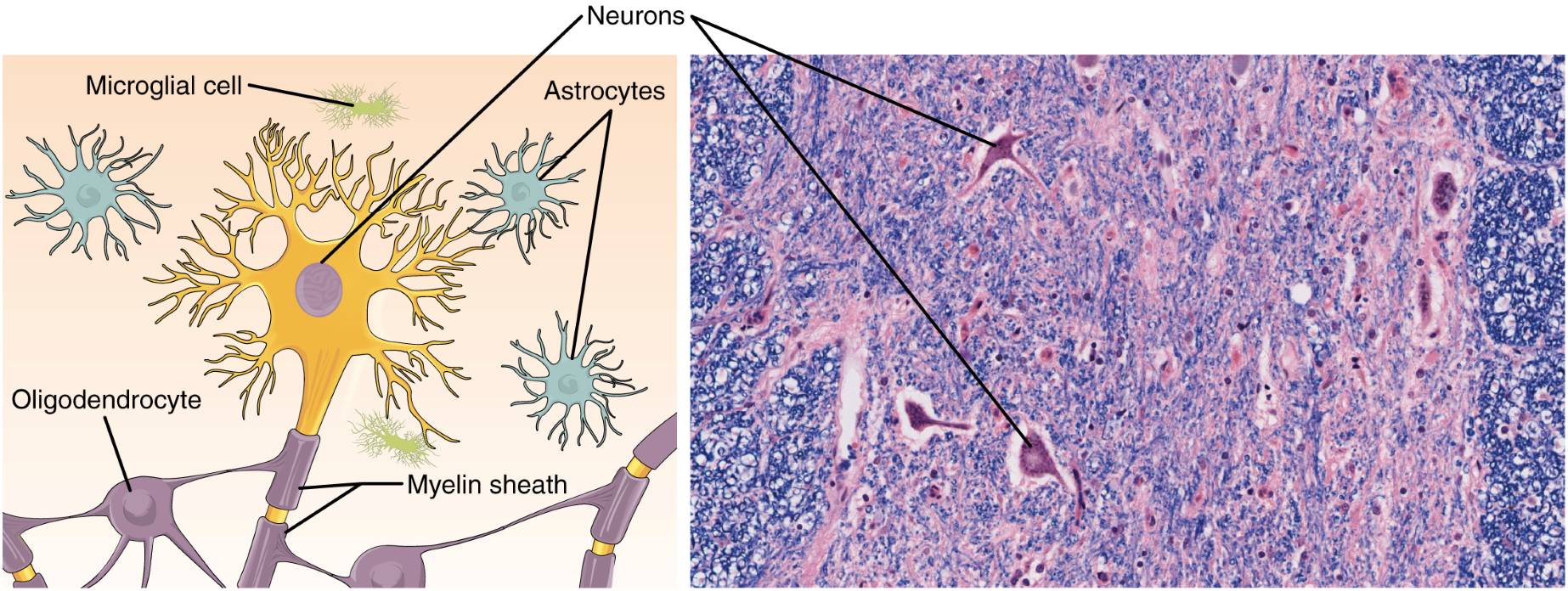
Nervous tissue forms the foundation of the nervous system, enabling the transmission and processing of signals throughout the body. This article explores the anatomical structure of nervous tissue, focusing on its cellular components—neurons and neuroglia—such as astrocytes, oligodendrocytes, and microglial cells, as illustrated in a detailed diagram and a micrograph at 872x magnification. By examining the structure and physical characteristics of nervous tissue, we uncover its critical role in coordinating bodily functions, from sensory perception to motor responses.
Labeled Anatomical Features of Nervous Tissue
Neurons
Neurons are the primary cells of nervous tissue, specialized to transmit electrical and chemical signals. They consist of a cell body, dendrites, and an axon, enabling communication across the nervous system.
Astrocytes
Astrocytes are star-shaped neuroglial cells that provide structural and metabolic support to neurons. They regulate the extracellular environment, maintain the blood-brain barrier, and facilitate nutrient delivery to neurons.
Microglial cell
Microglial cells are the immune cells of the nervous system, acting as macrophages to remove debris and pathogens. They play a key role in protecting nervous tissue by responding to injury and infection.
Oligodendrocyte
Oligodendrocytes are neuroglial cells responsible for producing the myelin sheath around axons in the central nervous system. This insulation enhances the speed of signal transmission along neurons.
Myelin sheath
The myelin sheath is a lipid-rich layer wrapped around axons by oligodendrocytes, insulating them to prevent signal loss. It allows for faster signal conduction through a process called saltatory conduction.
Overview of Nervous Tissue Composition
Nervous tissue is a complex network of cells working together to transmit and process information. Its primary components, neurons and neuroglia, each play distinct roles in maintaining neural function.
- Neurons are responsible for generating and transmitting action potentials, enabling rapid communication.
- Neuroglia, including astrocytes, oligodendrocytes, and microglial cells, support and protect neurons.
- The tissue is found in the brain, spinal cord, and peripheral nerves, forming the nervous system.
- Neurons vary in shape and size, depending on their specific function, such as sensory or motor roles.
- Neuroglia outnumber neurons and are essential for maintaining the neural environment.
- Nervous tissue is highly specialized, with a structure optimized for signal transmission.
This composition allows nervous tissue to coordinate complex bodily functions. From reflex actions to cognitive processes, its role is indispensable in daily life.
Physical Characteristics of Nervous Tissue
The physical properties of nervous tissue reflect its specialized role in signal transmission. These characteristics are crucial for its efficiency and functionality.
- Neurons have elongated axons, some extending over a meter, to connect distant parts of the body.
- The myelin sheath gives axons a white appearance and enhances signal speed by insulating them.
- Astrocytes have a star-like shape, maximizing their ability to interact with neurons and blood vessels.
- Microglial cells are smaller and more mobile, allowing them to patrol and respond to neural damage.
- Oligodendrocytes extend multiple processes to myelinate several axons simultaneously.
- Nervous tissue is soft and delicate, requiring protection by structures like the skull and meninges.
These physical traits highlight the tissue’s adaptation for rapid communication. They also underscore the importance of structural integrity in neural function.
Microscopic Insights into Nervous Tissue
The micrograph at 872x magnification reveals the cellular details of nervous tissue. This high-resolution view provides a deeper understanding of its organization.
- Astrocytes are visible with their characteristic star-shaped processes, interacting with neurons.
- Neurons stand out with their large cell bodies and darkly stained nuclei, indicating high metabolic activity.
- The myelin sheath appears as a clear area around axons, reflecting its insulating role.
- Microglial cells are less prominent but can be identified by their small, irregular shapes.
- The dense network of cells and processes illustrates the complexity of neural interactions.
- Staining techniques highlight the contrast between neurons and neuroglia, aiding in their identification.
Microscopic examination is essential for studying nervous tissue anatomy. It reveals the intricate relationships between its cellular components.
Functional Roles of Neurons and Neuroglia
Neurons and neuroglia work together to ensure the nervous system operates effectively. Their complementary roles are critical for signal transmission and neural health.
- Neurons generate action potentials through changes in membrane potential, driven by ion channels.
- Astrocytes regulate neurotransmitter levels, such as glutamate, to prevent excitotoxicity.
- Oligodendrocytes myelinate axons, increasing the speed of signal transmission via saltatory conduction.
- Microglial cells remove damaged cells and pathogens, maintaining a healthy neural environment.
- Neuroglia provide structural support, anchoring neurons in place within the tissue.
- Neurons form synapses, where neurotransmitters like dopamine facilitate signal transfer.
This teamwork ensures the nervous system can respond to stimuli efficiently. It also supports the maintenance of neural networks over time.
Clinical Relevance of Nervous Tissue
Nervous tissue health is vital for overall bodily function, and its dysfunction can lead to significant disorders. Understanding its structure aids in addressing neural conditions.
- Damage to the myelin sheath, as in multiple sclerosis, slows signal transmission and impairs motor function.
- Astrocyte dysfunction can disrupt the blood-brain barrier, leading to neural inflammation.
- Microglial overactivation is implicated in neurodegenerative diseases like Alzheimer’s, causing excessive inflammation.
- Oligodendrocyte loss can result in demyelination, affecting signal speed and coordination.
- Neuronal damage, such as in stroke, disrupts communication and leads to functional deficits.
- Protecting nervous tissue through a healthy lifestyle, including proper nutrition, supports neural function.
The clinical significance of nervous tissue underscores the need for ongoing research. Advances in neuroscience continue to improve our understanding and treatment of neural disorders.
Nervous tissue is a remarkable component of the body, orchestrating communication and coordination through its specialized cells. By exploring its anatomical structure and physical characteristics, we gain a deeper appreciation for its role in health and disease. This knowledge not only enhances our understanding of the nervous system but also informs strategies for maintaining its integrity.

