Explore the intricate superficial anatomy of the heart from a posterior perspective, revealing major blood vessels, coronary circulation, and key structures. This detailed view is essential for understanding the heart’s external features and its vascular supply, offering crucial insights into its overall function. A comprehensive grasp of this posterior anatomy is vital for diagnosing cardiovascular conditions and planning medical interventions.
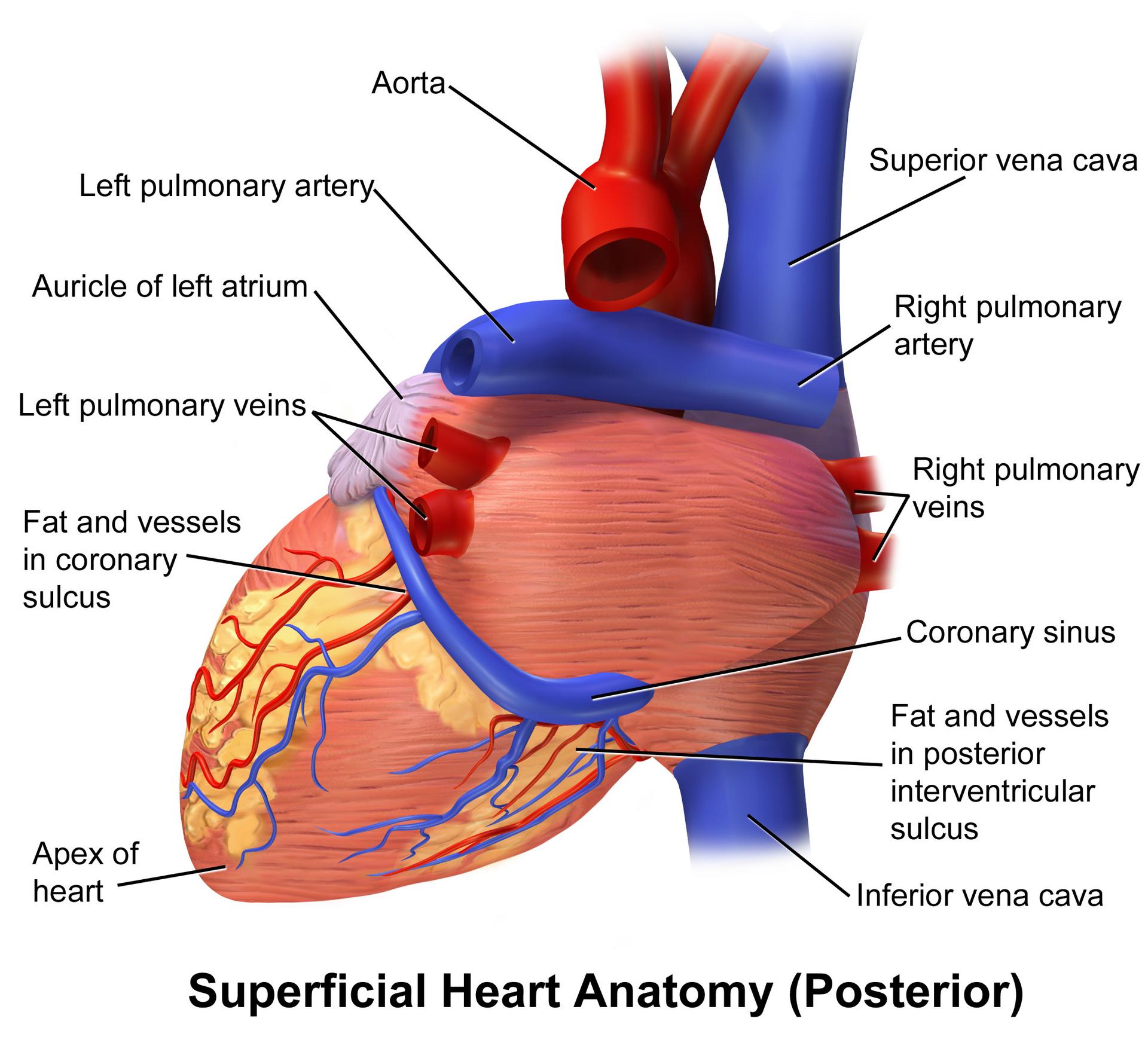
Aorta: The aorta is the largest artery in the human body, originating from the left ventricle and arching over the pulmonary artery. From this posterior view, its significant size and the beginning of its branching can be observed, highlighting its role in distributing oxygenated blood throughout the systemic circulation.
Superior vena cava: The superior vena cava is a large vein that carries deoxygenated blood from the upper half of the body, including the head, neck, and upper limbs, to the right atrium of the heart. Its prominent position posterior to the aorta indicates its crucial role in systemic venous return.
Left pulmonary artery: The left pulmonary artery branches from the main pulmonary artery and carries deoxygenated blood to the left lung. Its posterior location, along with its counterpart, underscores the heart’s connection to the pulmonary circulation for gas exchange.
Auricle of left atrium: The auricle of the left atrium is a small, ear-shaped muscular pouch that extends from the left atrium. While the left atrium receives oxygenated blood from the lungs, the auricle plays a role in increasing the capacity of the atrium and is a common site for thrombus formation in conditions like atrial fibrillation.
Right pulmonary artery: The right pulmonary artery branches from the main pulmonary artery and transports deoxygenated blood to the right lung. Both pulmonary arteries are integral to the pulmonary circulation, facilitating the re-oxygenation of blood.
Left pulmonary veins: The left pulmonary veins typically consist of two veins that carry oxygenated blood from the left lung back to the left atrium of the heart. Their entry into the left atrium from the posterior aspect is clearly visible.
Right pulmonary veins: The right pulmonary veins usually comprise two veins that carry oxygenated blood from the right lung to the left atrium. Along with the left pulmonary veins, they complete the pulmonary circuit, delivering oxygen-rich blood for systemic distribution.
Fat and vessels in coronary sulcus: The coronary sulcus, also known as the atrioventricular groove, encircles the heart and separates the atria from the ventricles. It houses major coronary arteries and veins, embedded in a layer of epicardial fat, which supply blood to the heart muscle itself.
Coronary sinus: The coronary sinus is a large venous structure located in the posterior aspect of the coronary sulcus, which collects most of the deoxygenated blood from the myocardium. It then drains this blood directly into the right atrium.
Fat and vessels in posterior interventricular sulcus: The posterior interventricular sulcus is a groove on the posterior surface of the heart that separates the right and left ventricles. It also contains important coronary vessels, including the posterior interventricular artery and middle cardiac vein, embedded in fat.
Apex of heart: The apex of the heart is the pointed, inferior portion of the organ, primarily formed by the left ventricle. From this posterior view, its general direction can be observed, though it points anteriorly and to the left.
Inferior vena cava: The inferior vena cava is a large vein that carries deoxygenated blood from the lower half of the body, including the trunk, abdomen, and lower limbs, to the right atrium. Its entry into the heart from the inferior and posterior aspect is a key feature of venous return.
The posterior view of the heart offers a unique perspective on its intricate vascular network and the pathways of blood returning to the heart. This anatomical illustration highlights the crucial role of the great vessels—the aorta, superior vena cava, and inferior vena cava—in systemic circulation, as well as the pulmonary arteries and veins connecting the heart to the lungs. It also prominently features the coronary circulation, demonstrating the vessels that nourish the heart muscle itself. Understanding these superficial details is fundamental for comprehending both normal cardiac function and the pathogenesis of various heart diseases.
A key feature revealed in this posterior view is the extensive network of coronary arteries and veins, which lie within the coronary sulcus and the interventricular sulci, often surrounded by adipose tissue. These vessels are responsible for supplying oxygenated blood to the myocardium and removing deoxygenated blood. Blockages in these coronary arteries can lead to myocardial ischemia, a condition where heart muscle tissue doesn’t receive enough blood flow. If prolonged, this can result in a myocardial infarction, commonly known as a heart attack, where heart muscle cells die due to lack of oxygen.
The coronary sinus, visible in the posterior coronary sulcus, is a major component of the heart’s venous drainage system, collecting deoxygenated blood from the cardiac veins and emptying it into the right atrium. Dysfunction of the coronary circulation, whether arterial or venous, can severely compromise cardiac function. For instance, atherosclerosis, the buildup of plaque in the arteries, is a leading cause of coronary artery disease, narrowing these vital vessels and reducing blood flow to the heart muscle.
- The coronary arteries are the first branches off the aorta.
- The heart receives its blood supply during diastole (relaxation).
- The left coronary artery typically branches into the anterior interventricular and circumflex arteries.
- The right coronary artery often supplies the SA and AV nodes.
This detailed posterior view is indispensable for cardiologists and cardiac surgeons, providing essential anatomical landmarks for diagnostic procedures and surgical interventions. It underscores the critical importance of a healthy coronary circulation for sustained cardiac performance and overall cardiovascular well-being. A thorough understanding of this superficial anatomy enhances our ability to prevent, diagnose, and treat the complex array of heart conditions that can arise.
This comprehensive posterior view of the superficial heart anatomy offers invaluable insights into the complex network of vessels and structures that sustain cardiac function. From the major arteries and veins facilitating global blood flow to the intricate coronary circulation nourishing the heart muscle itself, every detail contributes to the organ’s remarkable efficiency. A deep understanding of this anatomy is crucial for both medical professionals and individuals seeking to comprehend the intricate workings of the human heart and the origins of cardiovascular health and disease.

