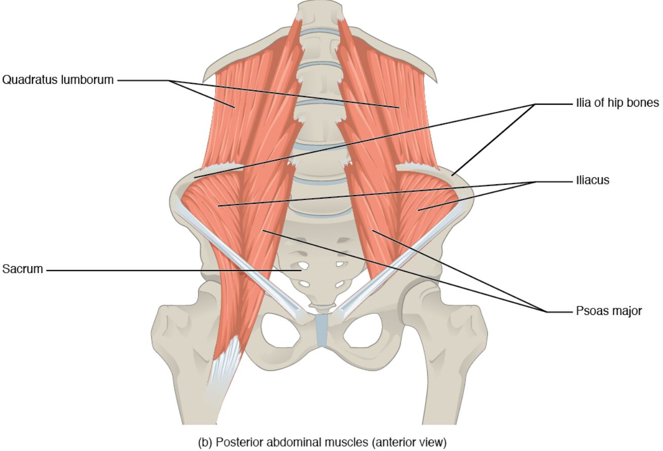
The posterior abdominal muscles play a vital role in supporting the lower back and facilitating movement of the lumbar spine and femur. This detailed examination of the posterior abdominal muscles in an anterior view provides a clear understanding of their anatomical structure and functional significance, essential for anyone exploring human physiology.
Labeled Parts Introduction
Quadratus lumborum
The quadratus lumborum is a deep muscle connecting the pelvis to the lumbar vertebrae and lower ribs, primarily responsible for lateral flexion of the spine and stabilizing the pelvis during locomotion. It also aids in extending the lumbar spine, contributing to overall posture.
Ilium of hip bones
The ilium of hip bones forms the broad, upper portion of the pelvis, serving as an attachment site for muscles like the iliacus and quadratus lumborum, and supports the upper body’s weight during sitting or standing. It is a key structural element of the pelvic girdle.
Iliacus
The iliacus is a flat, triangular muscle located in the iliac fossa of the pelvis, working with the psoas major to flex the hip joint, and is crucial for movements such as walking and climbing. It provides stability to the pelvis during weight-bearing activities.
Psoas major
The psoas major is a long muscle originating from the lumbar spine, extending through the pelvis to the femur, and is primarily responsible for flexing the hip joint and stabilizing the lower back. It collaborates with the iliacus to support leg movement and maintain posture.
Sacrum
The sacrum is a triangular bone at the base of the spine, formed by the fusion of sacral vertebrae, and serves as a foundation for the pelvic girdle while transmitting weight from the upper body to the lower limbs. It provides attachment points for ligaments and muscles, enhancing spinal support.
Overview of Posterior Abdominal Muscle Anatomy
The posterior abdominal muscles are essential for maintaining the stability of the lumbar spine and facilitating hip movements, as depicted in this anterior view. These muscles, including the quadratus lumborum and psoas major, work together to support the body’s core and enable a range of physical activities. Their intricate connections with the pelvis and spine highlight their importance in human anatomy.
- Forms a critical link between the spine and pelvis, ensuring coordinated movement.
- Protects the lower back by stabilizing the lumbar region during dynamic activities.
Structure of Posterior Abdominal Muscles
This image showcases the quadratus lumborum, a deep muscle flanking the lumbar spine, and the iliacus, which originates from the ilium of hip bones. The psoas major extends from the lumbar vertebrae to the femur, while the sacrum anchors the pelvic structure, illustrating a robust musculoskeletal framework.
- The quadratus lumborum is often evaluated for its role in lateral spine stability.
- The ilium of hip bones serves as a sturdy base for muscle attachments.
Role in Lumbar Spine and Hip Movement
The sacrum acts as a stable base, distributing weight between the spine and pelvis, while the psoas major and iliacus drive hip flexion essential for locomotion. The quadratus lumborum supports lateral flexion and spinal extension, playing a key role in maintaining posture and balance during movement.
- A strong psoas major enhances hip mobility for activities like running.
- The iliacus contributes to pelvic stability during weight-bearing tasks.
Clinical Relevance and Physical Health
The quadratus lumborum is often implicated in lower back pain due to its role in spinal stability, making it a focus in clinical assessments. The psoas major and iliacus can become tight, leading to anterior pelvic tilt, which may require stretching or strengthening exercises to restore balance.
- Weakness in the quadratus lumborum may lead to scoliosis or lumbar strain.
- The ilium of hip bones is a landmark for diagnosing pelvic misalignment.
Physical Examination and Rehabilitation
During physical exams, palpating the sacrum and ilium of hip bones helps assess pelvic alignment, while the psoas major is checked for tightness or weakness. Rehabilitation programs often target the iliacus and quadratus lumborum to alleviate back pain and improve core strength.
- Proper alignment of the sacrum is crucial for spinal health.
- Strengthening the psoas major can correct hip flexor imbalances.
Conclusion
The posterior abdominal muscles, as illustrated in this anterior view, form a complex system that supports the lumbar spine, pelvis, and hip joints with precision. Muscles like the quadratus lumborum, psoas major, and iliacus, anchored by the sacrum and ilium of hip bones, are vital for movement and stability. A thorough understanding of their anatomy enhances the ability to address musculoskeletal issues and optimize physical performance.

