The optic nerve and optic tract are critical components of the visual system, connecting the eye to the brain and facilitating the journey of visual information. This detailed anatomical drawing illustrates the pathway from the retina through the optic chiasm to the brain, highlighting the transition from peripheral to central structures. Understanding these neural connections enhances insight into how sight is processed and perceived within the cerebral anatomy.
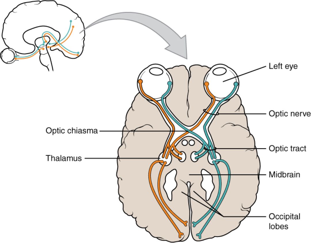
Labeled Components of the Visual Pathway
Optic nerve: The optic nerve extends from the retina at the back of the eye, carrying visual information via axons of ganglion cells. It transmits these signals to the optic chiasm, marking the beginning of the central visual pathway.
Optic chiasm: The optic chiasm is where the optic nerves partially cross, allowing visual fields from both eyes to be combined. This crossing ensures binocular vision and depth perception by merging input from the nasal and temporal retina.
Optic tract: The optic tract continues from the optic chiasm, carrying processed visual signals toward the brain’s thalamus and beyond. It contains axons that have crossed at the chiasm, relaying information to higher visual centers.
Eye: The eye serves as the initial receptor, capturing light through the retina where photoreceptors convert it into electrical signals. These signals initiate the visual pathway, traveling through the optic nerve.
Retina: The retina contains photoreceptors (rods and cones) that detect light and initiate visual processing. It sends this information via ganglion cell axons into the optic nerve.
Thalamus: The thalamus, specifically the lateral geniculate nucleus, receives signals from the optic tract and relays them to the visual cortex. It acts as a processing hub, refining visual data before further interpretation.
Midbrain: The midbrain, including the superior colliculus, receives some optic tract fibers for reflex actions like eye movement. It contributes to coordinating visual attention and orientation.
Occipital lobe: The occipital lobe houses the visual cortex, where processed visual information is interpreted to form coherent images. It integrates inputs from both optic tracts for complete visual perception.
Anatomy of the Optic Nerve
The optic nerve is the starting point of the visual pathway. This section explores its structure and function.
- Retinal Origin: The optic nerve begins at the optic disc, where ganglion cell axons exit the retina. This area lacks photoreceptors, creating the blind spot.
- Cranial Nerve II: Classified as cranial nerve II, the optic nerve carries approximately 1.2 million axons. It is sheathed in meninges, similar to the brain, reflecting its central nervous system status.
- Myelin Sheath: The nerve is myelinated by oligodendrocytes, enhancing signal speed to the chiasm. This insulation is crucial for efficient visual transmission.
- Pathway Protection: Enclosed in the optic foramen, the nerve is protected as it travels to the brain. Any compression here can lead to vision loss.
Structure and Role of the Optic Tract
The optic tract extends the visual pathway into the brain. This section details its anatomy and significance.
- Chiasm Transition: After the optic chiasm, the optic tract forms, carrying crossed and uncrossed retinal fibers. This structure ensures both hemifields are represented in each brain hemisphere.
- Lateral Geniculate Nucleus: The tract terminates in the lateral geniculate nucleus of the thalamus, a key relay station. This nucleus organizes visual input by layers corresponding to eye origin.
- Optic Radiation: Fibers from the thalamus form the optic radiation, projecting to the occipital lobe. This pathway preserves spatial organization of the visual field.
- Bilateral Representation: The optic tract’s design allows each hemisphere to process input from both eyes, supporting binocular vision. This integration is vital for depth perception.
Visual Processing in the Brain
The brain transforms raw visual data into meaningful perception. This section examines the higher visual centers.
- Thalamic Processing: The lateral geniculate nucleus filters and organizes visual signals before sending them to the cortex. It also receives feedback to adjust sensitivity.
- Occipital Cortex Function: The primary visual cortex (V1) in the occipital lobe maps the visual field topographically. Adjacent areas process color, motion, and form.
- Midbrain Contribution: The superior colliculus in the midbrain directs reflexive eye movements toward stimuli. It works with cortical areas for coordinated vision.
- Cortical Integration: Higher cortical areas, like V2 and V3, refine perception, combining inputs for object recognition. This multistage process underlies complex visual tasks.
Clinical Relevance of the Optic Pathway
Knowledge of the optic nerve and tract aids in diagnosing visual disorders. This section highlights clinical applications.
- Optic Neuritis: Inflammation of the optic nerve can cause pain and vision loss, often linked to multiple sclerosis. MRI can detect lesions along its course.
- Chiasmal Compression: Tumors like pituitary adenomas can press on the optic chiasm, leading to bitemporal hemianopia. Surgical intervention may be required.
- Tract Lesions: Damage to the optic tract may result in homonymous hemianopia, affecting half the visual field. Localization depends on the lesion’s side.
- Diagnostic Imaging: MRI and visual field testing map pathway integrity, guiding treatment. Early detection improves outcomes in neurological conditions.
The optic nerve and optic tract form a remarkable pathway that bridges the eye and brain, enabling the gift of sight. From the retina’s light capture to the occipital lobe’s image formation, this system exemplifies the intricacy of cerebral anatomy. Exploring these structures not only deepens appreciation for visual processing but also underscores the importance of protecting this pathway from injury or disease, paving the way for advances in neurology and vision care.
Additional SEO Titles:
- Optic Nerve and Tract: Understanding the Visual Pathway
- Exploring Optic Nerve Anatomy and Its Brain Connection
- Optic Tract and Chiasm: A Guide to Vision Processing
- Visual System Anatomy: From Eye to Occipital Lobe
- The Role of the Optic Nerve in Sight and Perception

