The human body’s range of motion extends beyond basic movements, enabled by the dynamic capabilities of synovial joints across various regions. This diagram details advanced movements such as supination, pronation, dorsiflexion, plantar flexion, eversion, inversion, protraction, retraction, depression, elevation, opposition, and reposition, illustrating their roles in everyday activities and joint function. Exploring this image provides a thorough understanding of the anatomical mechanisms that support the body’s diverse mobility.
Labelled Parts Explanation
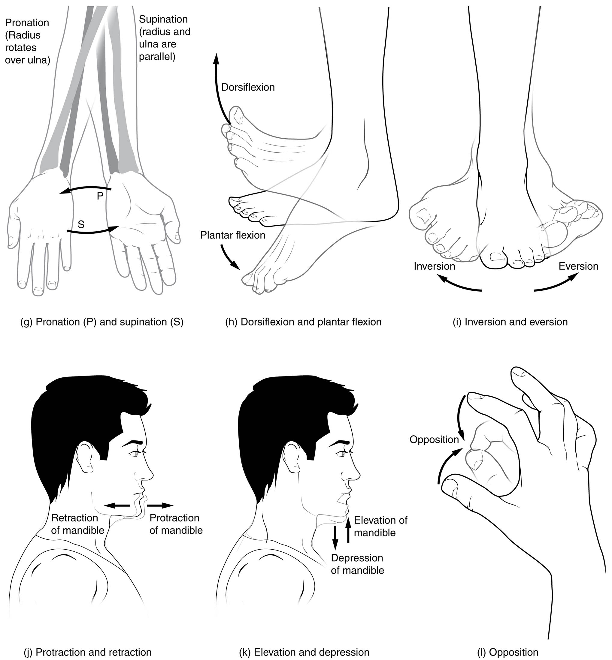
- Supination The supination movement turns the forearm so the palm faces forward or upward, with the radius and ulna aligning parallel. It is essential for actions like holding a bowl or turning a doorknob, facilitated by supinator muscles.
- Pronation The pronation movement rotates the forearm so the palm faces backward or downward, with the radius crossing over the ulna to form an “X” shape. It supports tasks like pouring water or pushing down, driven by pronator muscles.
- Dorsiflexion The dorsiflexion movement lifts the top of the foot toward the shin at the ankle joint, reducing the angle between them. It is crucial for walking uphill or lifting the foot during the swing phase, aided by tibialis anterior.
- Plantar flexion The plantar flexion movement extends the foot downward, pointing the toes and lifting the heel, increasing the ankle angle. It is vital for actions like standing on tiptoes or pushing off during walking, supported by gastrocnemius and soleus.
- Eversion The eversion movement turns the sole of the foot away from the body’s midline, as in tilting the foot outward. It assists in balancing on uneven surfaces, facilitated by peroneal muscles.
- Inversion The inversion movement rotates the sole of the foot toward the body’s midline, turning it inward. It helps with stability on sloped terrain, driven by tibialis posterior and other invertor muscles.
- Protraction The protraction movement pushes the mandible or other structures forward, such as jutting the chin out. It is used in chewing or speaking, supported by protractor muscles like the lateral pterygoid.
- Retraction The retraction movement pulls the mandible or structures back toward their original position, such as tucking the chin in. It aids in closing the jaw or aligning the head, facilitated by retractor muscles.
- Depression The depression movement lowers the mandible, opening the mouth by dropping the jaw downward. It is essential for yawning or biting into food, driven by depressor muscles like the digastric.
- Elevation The elevation movement raises the mandible, closing the mouth by lifting the jaw upward. It supports actions like chewing or clenching teeth, aided by elevator muscles like the masseter.
- Opposition The opposition movement brings the thumb’s tip into contact with the fingertips of the same hand, enabling a pincer grip. It is crucial for grasping objects, facilitated by the opponens pollicis.
- Reposition The reposition movement returns the thumb to its neutral position next to the index finger after opposition. It restores the hand’s resting alignment, supported by repositioning muscles.
Anatomical Overview of Advanced Body Movements
Synovial joints enable a wide array of advanced movements, each serving specific functional purposes. This diagram highlights the diversity of motion across the body’s joints.
- The supination and pronation rotate the forearm, affecting hand orientation.
- The dorsiflexion and plantar flexion control ankle and foot positioning.
- The eversion and inversion stabilize the foot on varied surfaces.
- The protraction, retraction, depression, elevation, opposition, and reposition govern jaw and thumb movements.
These actions reflect the joints’ adaptability to complex tasks.
Supination and Pronation of the Forearm
Supination and pronation allow rotational forearm movements. Their mechanics support hand functionality.
- The supination aligns the radius and ulna for a palm-up position.
- The pronation crosses the radius over the ulna for a palm-down stance.
- These motions occur at the radioulnar joints, aided by supinator and pronator muscles.
- They are essential for tasks requiring hand rotation.
This rotation enhances grip versatility.
Dorsiflexion and Plantar Flexion at the Ankle
Dorsiflexion and plantar flexion regulate foot movement. Their roles are key to locomotion.
- The dorsiflexion lifts the foot toward the shin, aiding in stepping.
- The plantar flexion points the toes, propelling the body forward.
- These movements occur at the ankle joint, driven by anterior and posterior leg muscles.
- They support walking, running, and standing balance.
This motion is critical for gait.
Eversion and Inversion of the Foot
Eversion and inversion provide lateral foot stability. Their function aids in uneven terrain navigation.
- The eversion turns the sole outward, enhancing lateral balance.
- The inversion rotates the sole inward, supporting medial stability.
- These occur at the subtalar and midtarsal joints, powered by peroneal and posterior muscles.
- They prevent ankle sprains during movement.
This stability is vital for foot health.
Protraction and Retraction of the Mandible
Protraction and retraction control jaw positioning. Their actions support oral functions.
- The protraction moves the chin forward, aiding in biting.
- The retraction pulls the chin back, aligning the jaw.
- These occur at the temporomandibular joint, driven by pterygoid muscles.
- They facilitate chewing and speech.
This motion enhances jaw mobility.
Depression and Elevation of the Mandible
Depression and elevation manage mouth opening and closing. Their coordination supports feeding.
- The depression lowers the jaw, opening the mouth for intake.
- The elevation raises the jaw, closing the mouth for chewing.
- These occur at the temporomandibular joint, powered by digastric and masseter muscles.
- They are essential for mastication and swallowing.
This action is key to oral health.
Opposition and Reposition of the Thumb
Opposition and reposition enable thumb dexterity. Their function supports hand precision.
- The opposition allows the thumb to touch fingertips, enabling grip.
- The reposition returns the thumb to its neutral alignment.
- These occur at the carpometacarpal joint, driven by opponens pollicis.
- They are crucial for fine motor skills like writing.
This movement enhances hand function.
Physiological Importance of Body Movements
These movements are integral to maintaining physical functionality. Their coordination supports daily life.
- The supination and pronation aid in hand manipulation.
- The dorsiflexion and plantar flexion ensure gait stability.
- The eversion, inversion, protraction, retraction, depression, elevation, opposition, and reposition enhance balance and dexterity.
- Joint health sustains these motions over time.
This versatility underpins an active lifestyle.
Clinical Relevance of Joint Movements
Understanding these movements assists in diagnosing musculoskeletal conditions. These actions are key clinical indicators.
- Limited supination or pronation may suggest radioulnar joint injury.
- Abnormal dorsiflexion or plantar flexion can indicate ankle disorders.
- Reduced opposition might reflect thumb arthritis.
- Therapy targets these movements to restore function.
This knowledge guides rehabilitation efforts.
Conclusion
The movements of the body medical description diagram 2 provides a detailed exploration of advanced motions, including supination, pronation, dorsiflexion, plantar flexion, eversion, inversion, protraction, retraction, depression, elevation, opposition, and reposition, facilitated by synovial joints. By examining how these movements enhance mobility across the forearm, ankle, foot, jaw, and thumb, one gains insight into the body’s anatomical versatility. This understanding serves as a foundation for studying musculoskeletal health and addressing related concerns, encouraging further exploration of joint mechanics and their role in supporting an active life.

