Histological Structure of Endometrial Tissue: A Microscopic Analysis

Date:

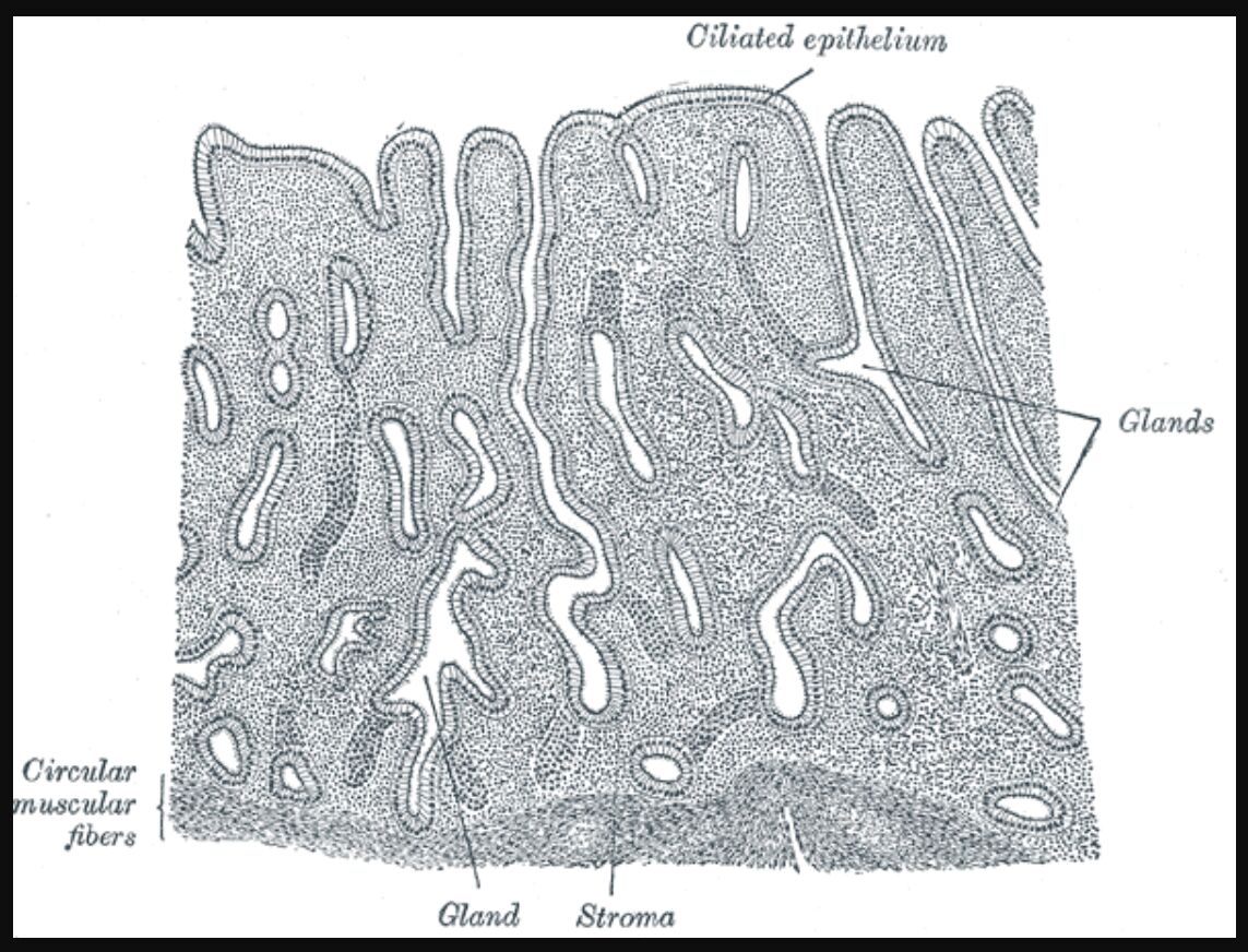
The endometrial tissue represents one of the most dynamic and specialized epithelial surfaces in the human body, demonstrating remarkable cyclical changes in response to hormonal fluctuations. This microscopic examination reveals the intricate architecture of the endometrial lining, highlighting its essential components and their roles in reproductive function.

vertical-section-of-mucous-membrane-of-human-uterus Histological Structure of Endometrial Tissue: A Microscopic Analysis

By Henry Vandyke CarterHenry Gray (1918) Anatomy of the Human Body (See “Book” section below)Bartleby.com: Gray’s Anatomy, Plate 1169, Public Domain, Link

Ciliated epithelium The ciliated epithelium forms the luminal surface of the endometrium. These specialized cells contain motile cilia that help move secretions and potentially assist in gamete transport, while also providing a protective barrier against pathogens.

- Advertisement -

Recommended Study Resource

Gray's Anatomy: The Anatomical Basis of Clinical Practice

Enhance your anatomical knowledge with Gray's Anatomy: The Anatomical Basis of Clinical Practice. This authoritative text offers in-depth insights and illustrations, perfect for medical students and practitioners aiming for clinical excellence.

Shop Now on Amazon

At AnatomyNote.com, we offer free resources on anatomy, pathology, and pediatric medicine for medical students and professionals. Purchasing through our Amazon links, like Gray's Anatomy, supports our server costs and content creation at no additional cost to you.

Disclosure: As an Amazon Associate, we earn a commission from qualifying purchases.

Disclosure: As an Amazon Associate, we earn a commission from qualifying purchases at no extra cost to you.

Glands The endometrial glands are tubular structures that extend from the surface epithelium into the underlying stroma. These glands undergo cyclical changes in morphology and secretory activity throughout the menstrual cycle, producing substances essential for implantation and early embryo development.

Circular muscular fibers The circular muscular fibers form part of the myometrial layer beneath the endometrium. These smooth muscle bundles are arranged in a circular pattern and play crucial roles in uterine contractility and vascular regulation.

Gland Individual endometrial glands demonstrate distinct morphological features with columnar epithelial cells. These structures respond to hormonal signals and produce glycogen-rich secretions during the secretory phase of the menstrual cycle.

Stroma The endometrial stroma consists of specialized connective tissue that supports the glands and blood vessels. This tissue undergoes significant remodeling during the menstrual cycle and decidualization during pregnancy, with characteristic changes in cellular composition and extracellular matrix.

- Advertisement -

Anatomy Flash Cards

Master anatomy with detailed, exam-ready flash cards.

Buy on Amazon

AnatomyNote.com offers free anatomy and pathology resources. Your purchase of Anatomy Flash Cards supports our site at no extra cost.

As an Amazon Associate, we earn from qualifying purchases.

Understanding Endometrial Histology

Microscopic Architecture

The endometrial tissue demonstrates complex structural organization at the microscopic level. Each component plays a specific role in maintaining reproductive function and responding to hormonal signals. The intricate arrangement of epithelial and stromal elements enables the endometrium to undergo dramatic cyclic changes.

Epithelial Components

Surface Epithelium

The luminal epithelium consists of two main cell types:

  • Ciliated cells (15-20% of surface cells)
  • Secretory cells (80-85% of surface cells)

These cells form a continuous barrier and undergo significant morphological changes throughout the menstrual cycle, varying in height from 10-20 μm.

Glandular Structure

The endometrial glands demonstrate:

  • Simple columnar epithelium
  • Basal lamina
  • Secretory apparatus
  • Stem cell populations

Stromal Organization

Cellular Components

The stromal compartment contains:

  • Fibroblast-like cells
  • Immune cells
  • Spiral arterioles
  • Extracellular matrix

Vascular Elements

The vascular network includes:

  • Spiral arteries
  • Capillary plexuses
  • Lymphatic vessels
  • Specialized sinusoids

Functional Significance

Cyclic Changes

The endometrium undergoes regular modifications:

  • Proliferative phase changes
  • Secretory transformation
  • Menstrual shedding
  • Regeneration

Clinical Implications

Understanding histological features aids in:

  • Pathological diagnosis
  • Treatment planning
  • Fertility assessment
  • Disease monitoring

Molecular Characteristics

Key molecular markers include:

  • Estrogen receptors (ERα and ERβ)
  • Progesterone receptors (PR-A and PR-B)
  • Cell adhesion molecules
  • Growth factors

Modern Applications

Advanced techniques enable:

  • 3D tissue reconstruction
  • Single-cell analysis
  • Molecular mapping
  • Real-time imaging
  1. Endometrial Histology: A Comprehensive Guide to Microscopic Structure
  2. Understanding Endometrial Tissue: From Cells to Function
  3. Microscopic Anatomy of the Endometrium: A Clinical Guide
  4. Endometrial Architecture: Histological Analysis for Medical Professionals
  5. Comprehensive Guide to Endometrial Tissue Structure and Function
Image source:

LEAVE A REPLY

Please enter your comment!
Please enter your name here

Share post:

Popular

spot_imgspot_img

Subscribe

More like this
Related

Exploring the Cardiac Muscle Anatomical Structure Diagram

The Cardiac Muscle Anatomical Structure Diagram offers a detailed glimpse into the intricate architecture of the heart's muscular tissue, essential for its rhythmic contractions. This image highlights key components such as intercalated discs, gap junctions, and desmosomes, which work together to ensure coordinated heart function. By examining this diagram, one can gain a deeper understanding of how cardiac muscle fibers support the continuous pumping action vital for circulation and overall health.

Epithelial Tissue Types: Structure, Location, and Function in Human Anatomy

Epithelial tissues are vital components of the human body, serving as protective barriers, facilitating absorption, and enabling secretion across various organs. This summary of epithelial tissue cells highlights their diverse types, including simple, stratified, pseudostratified, and transitional epithelia, each with specific locations and functions. From the lungs to the urinary tract, these tissues are uniquely adapted to their roles, ensuring physiological balance and organ functionality. This article provides a detailed exploration of epithelial tissue types, their anatomical locations, and their critical contributions to human anatomy.

Cell Anatomy from the 1918 Human Body Edition: A Historical Perspective

This image presents a detailed diagram of a cell as depicted in the 1918 edition of "Anatomy of the Human Body," offering a historical view of cellular structure based on early 20th-century understanding. It serves as an educational tool for medical students and professionals to appreciate the evolution of cellular anatomy knowledge. The following sections explore each labeled component, providing insights into their historical context and anatomical significance.

Exploring Types of Epithelial Tissue: Simple, Stratified, and Pseudostratified Epithelia

Epithelial tissues are fundamental to the human body, forming protective layers and linings that cover surfaces and cavities. These tissues are classified into simple, stratified, and pseudostratified types based on the number and arrangement of cell layers, each serving distinct anatomical and physiological roles. Comprising cells like squamous, cuboidal, and columnar, epithelial tissues are essential for functions such as absorption, secretion, and protection. This article delves into the structure and significance of these tissues, providing a detailed analysis of their labeled components and their critical roles in human anatomy.