The fetal circulatory system is a remarkably adapted network designed to support prenatal life, where the lungs are non-functional and nutrient/gas exchange occurs via the placenta. This intricate system includes several unique shunts that bypass the pulmonary circulation, ensuring that oxygenated blood and essential nutrients are efficiently delivered to the developing fetus. The provided diagram offers a comprehensive overview of this specialized circulation, detailing the major vessels, the role of the placenta, and the key shunts that reroute blood flow. Understanding the fetal circulation is crucial for appreciating how the fetus thrives in utero and the physiological transitions that occur at birth.
Key Components and Shunts of Fetal Circulation
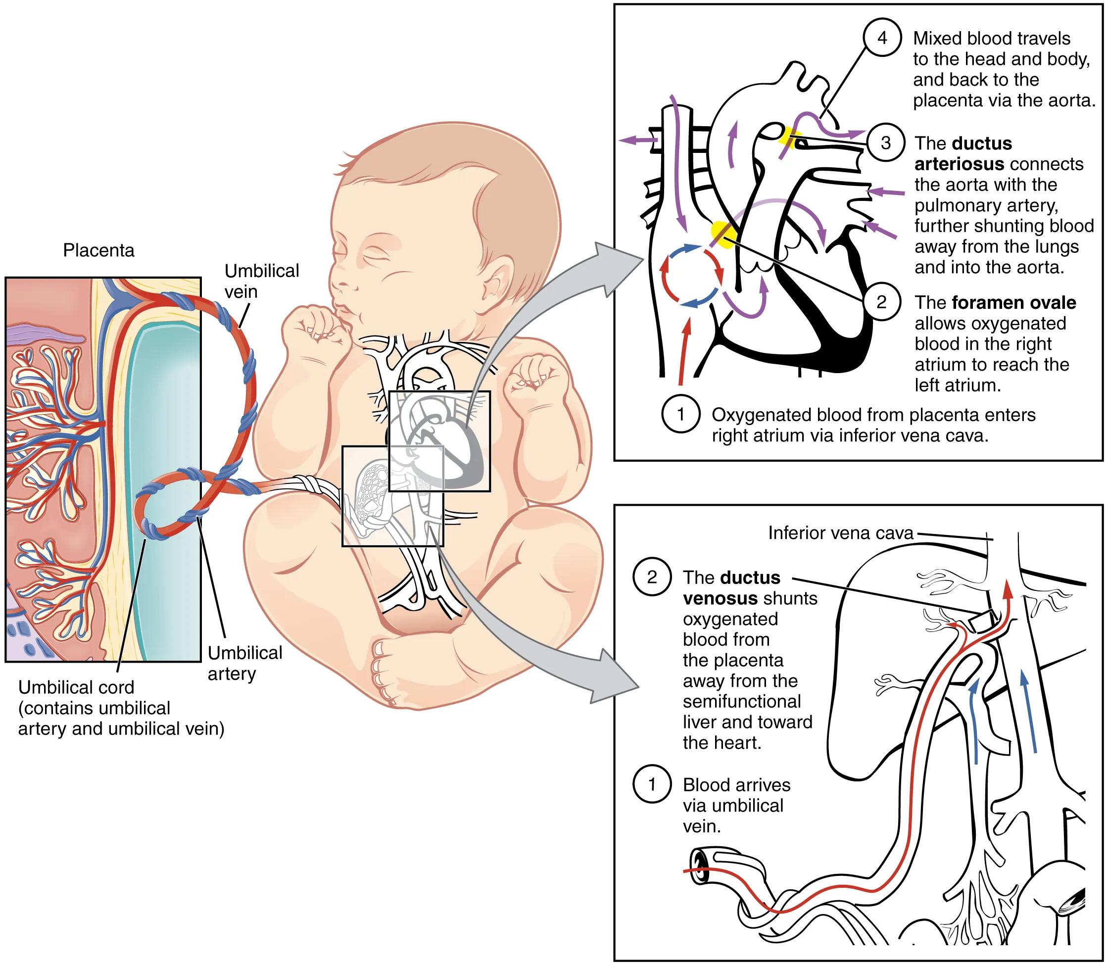
Placenta: The placenta is a vital organ during pregnancy, facilitating the exchange of oxygen, nutrients, and waste products between the mother and the fetus. It serves as the primary site for gas exchange, effectively acting as the “lungs” for the fetus.
Umbilical vein: This vessel carries oxygenated, nutrient-rich blood from the placenta to the fetus. It is a singular vein, distinct from the two umbilical arteries.
Umbilical artery: These arteries carry deoxygenated blood and metabolic waste products from the fetus back to the placenta for removal. There are typically two umbilical arteries.
Umbilical cord (contains umbilical artery and umbilical vein): The umbilical cord is the lifeline connecting the fetus to the placenta. It contains the umbilical vein and two umbilical arteries, encased in Wharton’s jelly for protection.
Ductus venosus: (1) Blood arrives via umbilical vein. (2) The ductus venosus shunts oxygenated blood from the placenta away from the semifunctional liver and toward the heart. This vascular shunt bypasses the liver sinusoids, allowing highly oxygenated blood to quickly reach the inferior vena cava and subsequently the right atrium.
Inferior vena cava: A large vein that carries deoxygenated blood from the lower and middle body to the right atrium of the heart. In fetal circulation, it also carries the highly oxygenated blood shunted through the ductus venosus.
Foramen ovale: (2) The foramen ovale allows oxygenated blood in the right atrium to reach the left atrium. This opening between the right and left atria shunts a significant portion of oxygenated blood directly into the systemic circulation, bypassing the pulmonary circuit.
Ductus arteriosus: (3) The ductus arteriosus connects the aorta with the pulmonary artery, further shunting blood away from the lungs and into the aorta. This shunt diverts blood from the pulmonary artery directly into the aorta, bypassing the high-resistance pulmonary circulation.
Mixed blood travels to the head and body, and back to the placenta via the aorta: (4) This indicates that the blood delivered to the systemic circulation is a mixture of oxygenated blood from the placenta and deoxygenated blood from the fetal body. This mixed blood then circulates throughout the fetus and returns to the placenta for reoxygenation and waste removal.
The Unique Pathways of Fetal Blood Flow
In fetal life, the lungs are collapsed and filled with fluid, offering high resistance to blood flow, making them inefficient for gas exchange. To circumvent this, the fetal circulatory system employs specialized shunts. Oxygenated blood, rich in nutrients, arrives from the placenta via the umbilical vein. A significant portion of this blood bypasses the liver through the ductus venosus, directly entering the inferior vena cava. This ensures that the most highly oxygenated blood reaches the heart.
Upon entering the right atrium, the majority of this blood is shunted directly into the left atrium through the foramen ovale, an opening in the interatrial septum. From the left atrium, it flows into the left ventricle and then into the aorta, supplying the head and upper extremities with relatively well-oxygenated blood. This preferential flow is crucial for the development of the brain. The remaining blood in the right atrium mixes with deoxygenated blood from the fetal body and enters the right ventricle.
From the right ventricle, blood is pumped into the pulmonary artery. However, due to the high pulmonary vascular resistance, most of this blood is diverted away from the lungs by the ductus arteriosus, which connects the pulmonary artery directly to the aorta. This mixed blood then travels down the aorta, supplying the lower body. A portion of this blood is then returned to the placenta via the umbilical arteries for reoxygenation and waste removal.
- The efficiency of these shunts is paramount for fetal survival, as they ensure that vital organs receive sufficient oxygen and nutrients despite the non-functional lungs.
At birth, a dramatic transition occurs. With the first breaths, the lungs inflate, pulmonary vascular resistance drops, and the shunts (ductus venosus, foramen ovale, and ductus arteriosus) functionally close, redirecting blood flow to establish the adult circulatory pattern.
Conclusion
The fetal circulatory system is a masterpiece of physiological adaptation, uniquely designed to sustain life in the intrauterine environment. The diagram clearly illustrates the crucial roles of the placenta, the umbilical vessels, and the three major shunts—the ductus venosus, foramen ovale, and ductus arteriosus—in bypassing the non-functional fetal lungs. Understanding these specialized pathways is fundamental to appreciating the mechanisms of fetal oxygenation and nutrient delivery, as well as the remarkable cardiovascular transitions that occur immediately after birth to support independent extrauterine life.

