The male reproductive system is a complex network of organs and structures essential for reproduction and hormonal regulation, as depicted in the provided image. This article provides a comprehensive look at the anatomical components illustrated, offering insights into their functions and interconnections. By understanding this system’s layout, one can appreciate its critical role in overall health and fertility.
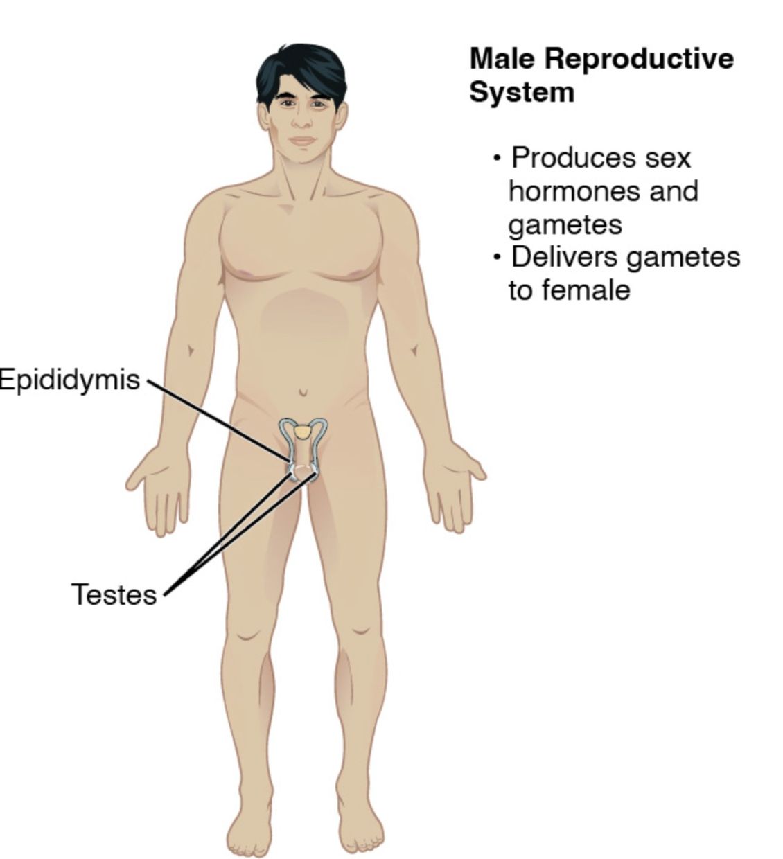
Testes: These are the primary male reproductive organs located in the scrotum, responsible for producing sperm and testosterone. Their dual role supports both reproduction and the development of secondary sexual characteristics.
Epididymis: This coiled tube sits atop each testis, where sperm mature and gain motility after being produced. It serves as a storage site before sperm are transported to the vas deferens.
Vas deferens: This long tube carries mature sperm from the epididymis to the urethra during ejaculation. It plays a key role in the transport mechanism of the reproductive process.
Prostate gland: Located below the bladder, this gland produces a fluid that forms part of semen, nourishing and protecting sperm. Its secretions are vital for sperm viability during ejaculation.
Seminal vesicles: These paired glands contribute a significant portion of seminal fluid, rich in fructose to energize sperm. They are situated behind the bladder and connect to the vas deferens.
Urethra: This tube extends from the bladder through the penis, serving as a conduit for both urine and semen. Its dual function is regulated by sphincter muscles to prevent mixing.
Penis: This external organ facilitates sexual intercourse and the expulsion of semen or urine. It contains erectile tissues that enable erection through blood flow.
Scrotum: This sac of skin and muscle houses the testes, maintaining an optimal temperature for sperm production. Its regulation is crucial for reproductive health.
Anatomical Structure of the Male Reproductive System
The male reproductive system’s anatomy is intricately designed for both reproduction and hormone production. Each component contributes to the system’s overall efficiency.
- The testes produce up to 1,500 sperm per second under hormonal influence.
- The epididymis can store sperm for several weeks if not ejaculated.
- The vas deferens contracts during ejaculation to propel sperm forward.
- The prostate gland enlarges with age, sometimes leading to benign prostatic hyperplasia.
- The seminal vesicles add volume and nutrients to semen, enhancing fertility.
Physiological Functions and Hormonal Regulation
The physiological roles of the male reproductive system are supported by hormonal interplay. This process ensures proper development and function.
- The testes release testosterone, driving puberty and maintaining libido.
- The epididymis matures sperm, preparing them for fertilization.
- The prostate gland secretes alkaline fluid to neutralize vaginal acidity.
- The seminal vesicles provide energy through fructose for sperm motility.
- The urethra and penis coordinate to deliver sperm during reproduction.
Clinical Relevance and Health Considerations
While the image focuses on anatomy, understanding the male reproductive system aids in recognizing potential health issues. Regular check-ups can prevent complications.
- The scrotum’s temperature regulation can be disrupted by varicocele, affecting fertility.
- Enlarged prostate gland may require monitoring for cancer risk.
- Blockages in the vas deferens can lead to infertility, treatable with surgery.
- Infections in the urethra or penis may cause urethritis, requiring antibiotics.
- Hormonal imbalances from the testes can impact overall well-being.
The male reproductive system, as illustrated, showcases the harmonious function of the testes, epididymis, vas deferens, prostate gland, seminal vesicles, urethra, penis, and scrotum in supporting reproduction. This anatomical overview provides a foundation for exploring its physiological roles and clinical significance. By studying these structures, one can better understand their importance in maintaining health and addressing reproductive challenges effectively.

