Coronary angiograms provide a critical view of atherosclerotic coronary arteries, revealing blockages that impede blood flow and oxygen delivery to the heart. This article explores the provided X-ray image, detailing how the dye highlights occluded arteries and the potential consequences, including ischemia, hypoxia, and myocardial infarction. Understanding these imaging findings can improve awareness and guide effective management of this serious cardiovascular condition.
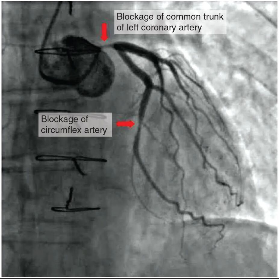
Occluded coronary artery 1: This label identifies the first blocked coronary artery, visible due to the contrast dye, indicating a significant buildup of atherosclerotic plaque. The blockage reduces blood flow, potentially leading to cardiac tissue damage if not addressed promptly.
Occluded coronary artery 2: This marks the second coronary artery affected by occlusion, similarly highlighted by the dye, showing another site of plaque accumulation. Its obstruction further compromises the heart’s oxygen supply, increasing the risk of a heart attack.
Contrast dye: This substance, injected during the angiogram, enhances the visibility of blood vessels on the X-ray. It outlines the occluded coronary arteries, allowing precise identification of the blockages.
Anatomy of Coronary Arteries
The coronary arteries are vital for supplying blood to the heart muscle. The angiogram reveals how atherosclerotic coronary arteries disrupt this essential function.
- The occluded coronary artery 1 and occluded coronary artery 2 are branches of the coronary circulation.
- These arteries normally deliver oxygen-rich blood to the myocardium.
- Atherosclerosis causes plaque buildup, narrowing the arterial lumen.
- The contrast dye fills the vessels, making blockages stand out clearly.
- This anatomical insight is crucial for planning interventions.
Pathophysiology of Atherosclerosis
Atherosclerotic coronary arteries lead to a cascade of physiological changes due to reduced blood flow. The angiogram illustrates the severity of this process.
- Plaque in occluded coronary artery 1 and occluded coronary artery 2 restricts blood flow, causing ischemia.
- Ischemia results in hypoxia, depriving cardiac tissue of adequate oxygen.
- Prolonged oxygen deprivation can lead to myocardial infarction, or heart muscle death.
- The contrast dye highlights the extent of narrowing, aiding diagnosis.
- Inflammatory processes within the arteries exacerbate plaque growth.
Clinical Management and Prognosis
Managing atherosclerotic coronary arteries requires a multifaceted approach to restore blood flow. The angiogram findings guide these critical decisions.
- Angioplasty or stenting can open occluded coronary artery 1 and occluded coronary artery 2.
- Medications like statins and antiplatelets reduce plaque progression and prevent clots.
- Bypass surgery may be considered for severe blockages shown by the contrast dye.
- Lifestyle changes, such as diet and exercise, support long-term vascular health.
- Prognosis improves with early intervention, depending on the occlusion’s extent.
The coronary angiogram offers a compelling view of atherosclerotic coronary arteries, with the contrast dye revealing the critical blockages in occluded coronary artery 1 and occluded coronary artery 2. These insights underscore the importance of timely diagnosis and treatment to prevent ischemia, hypoxia, and potentially fatal myocardial infarction. By leveraging such imaging, healthcare providers can tailor strategies to enhance patient outcomes and promote heart health.

