Delve into the intricate structures of the human mouth with this comprehensive guide, offering an anterior view. Learn about the lips, tongue, palate, gums, and teeth, and understand their vital roles in speech, taste, and the crucial first steps of digestion.
The human mouth is a complex and fascinating structure, serving as the gateway to the digestive and respiratory systems. From its role in articulating speech to initiating the mechanical and chemical breakdown of food, each component plays a vital role. This detailed exploration of the mouth from an anterior view highlights its anatomical features, providing insight into its multifaceted functions and overall importance to our health.
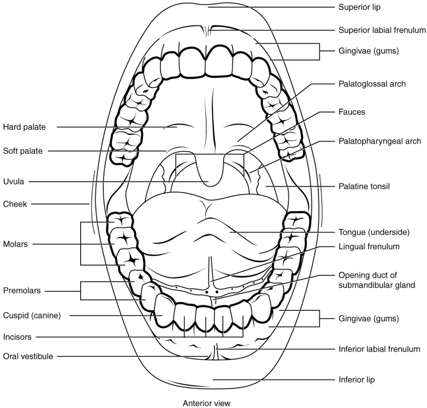
Superior lip: The superior lip, or upper lip, is a mobile, muscular fold that forms the upper boundary of the oral opening. It plays a crucial role in speech articulation, facial expressions, and containing food and liquids within the mouth.
Superior labial frenulum: This is a small fold of mucous membrane that connects the inner surface of the upper lip to the gum tissue between the upper central incisors. It provides stability to the upper lip and helps limit its movement.
Gingivae (gums): The gingivae, commonly known as gums, are the specialized mucosal tissue that surrounds the teeth and covers the alveolar bone. Healthy gums are pink, firm, and essential for supporting and protecting the teeth.
Palatoglossal arch: This arch is a fold of mucous membrane that extends from the soft palate to the side of the tongue. It forms the anterior boundary of the fauces, the opening into the oropharynx.
Fauces: The fauces represent the arched opening at the back of the mouth, leading into the pharynx. It is bordered by the palatoglossal and palatopharyngeal arches.
Palatopharyngeal arch: This fold of mucous membrane extends from the soft palate to the lateral wall of the pharynx, forming the posterior boundary of the fauces. It plays a role in swallowing by helping to close off the nasopharynx.
Palatine tonsil: Located between the palatoglossal and palatopharyngeal arches, the palatine tonsils are lymphoid tissues. They are part of the body’s immune system, helping to protect against infections entering through the mouth.
Tongue (underside): The underside of the tongue is covered by a thin, smooth mucous membrane. This area is rich in blood vessels, which can be easily seen.
Lingual frenulum: This fold of mucous membrane connects the underside of the tongue to the floor of the mouth. It helps anchor the tongue and can influence its range of motion.
Opening duct of submandibular gland: These small openings on either side of the lingual frenulum are where saliva produced by the submandibular glands is released into the mouth. Saliva is vital for digestion and oral hygiene.
Inferior labial frenulum: Similar to its superior counterpart, this fold of mucous membrane connects the inner surface of the lower lip to the gum tissue. It stabilizes the lower lip and restricts its excessive movement.
Inferior lip: The inferior lip, or lower lip, forms the lower boundary of the oral opening. It is equally important for speech, facial expressions, and retaining food and liquids in the mouth.
Hard palate: The hard palate forms the anterior, bony part of the roof of the mouth, separating the oral cavity from the nasal cavity. It provides a rigid surface against which the tongue can press food during chewing and swallowing.
Soft palate: The soft palate is the posterior, fleshy part of the roof of the mouth, composed of muscle and connective tissue. During swallowing, it elevates to close off the nasopharynx, preventing food from entering the nasal cavity.
Uvula: The uvula is a small, fleshy projection hanging from the middle of the soft palate. It assists in speech, particularly in forming certain sounds, and helps to seal off the nasal cavity during swallowing.
Cheek: The cheeks form the lateral walls of the oral cavity, composed primarily of buccinator muscles. They play a significant role in holding food between the teeth during mastication and in speech articulation.
Molars: Molars are the large, flat teeth located at the back of the mouth, designed for grinding and crushing food. Adults typically have twelve molars, including wisdom teeth.
Premolars: Premolars, also known as bicuspids, are located between the canines and molars. They have two cusps and are involved in both tearing and grinding food.
Cuspid (canine): The cuspids, or canines, are the pointed teeth located at the corners of the mouth. They are designed for tearing food and are often the longest teeth in the mouth.
Incisors: Incisors are the flat, chisel-shaped teeth at the front of the mouth. Their primary function is to cut and shear food into smaller pieces.
Oral vestibule: The oral vestibule is the space between the lips or cheeks and the teeth and gums. This area can be seen when the lips are pulled away from the teeth.
The oral cavity, commonly known as the mouth, is a multifaceted anatomical region that initiates the digestive process and plays a pivotal role in communication. Beyond merely housing the teeth, the mouth encompasses a rich tapestry of structures, each contributing to its remarkable functionality. It is where food intake, initial mechanical and chemical breakdown, taste perception, and the formation of speech sounds all converge. A healthy mouth is fundamental to overall systemic health, impacting nutrition, social interaction, and quality of life.
The mouth is generally understood to include several key components that work in concert:
- Lips: These muscular folds form the anterior boundary, crucial for speech, facial expression, and retaining food.
- Tongue: A highly muscular organ involved in taste, chewing, swallowing, and speech.
- Palate: Comprising the hard and soft sections, it forms the roof of the mouth, separating the oral cavity from the nasal cavity.
- Gums (Gingivae): The protective tissue surrounding the teeth, securing them in their sockets.
- Teeth: Essential for mastication, allowing for the mechanical breakdown of food.
This intricate interplay of structures ensures the mouth performs its diverse functions effectively. From the moment food enters, the lips seal the oral cavity, while the teeth begin the process of mastication. The tongue manipulates the food, mixing it with saliva, which not only lubricates but also begins chemical digestion through enzymes like salivary amylase. Simultaneously, the tongue and palate are critical for forming sounds, enabling clear speech. Understanding the precise anatomy and function of each part of the mouth provides a deeper appreciation for its significance in human physiology and daily life. Maintaining optimal oral hygiene and health is therefore not just about preventing cavities, but about preserving the integrity of this vital gateway to our bodies.

