Epithelial tissues are vital components of the human body, serving as protective barriers, facilitating absorption, and enabling secretion across various organs. This summary of epithelial tissue cells highlights their diverse types, including simple, stratified, pseudostratified, and transitional epithelia, each with specific locations and functions. From the lungs to the urinary tract, these tissues are uniquely adapted to their roles, ensuring physiological balance and organ functionality. This article provides a detailed exploration of epithelial tissue types, their anatomical locations, and their critical contributions to human anatomy.
Labeled Components of Epithelial Tissue Summary
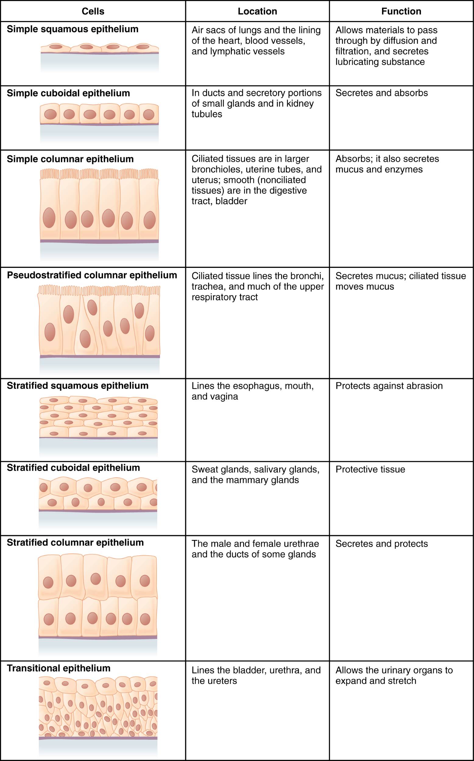
Simple Squamous Epithelium
Simple squamous epithelium consists of a single layer of flat cells, ideal for rapid diffusion and filtration. It is located in the air sacs of the lungs, the lining of the heart, blood vessels, and lymphatic vessels, where it allows materials to pass through efficiently.
Simple Cuboidal Epithelium
Simple cuboidal epithelium features a single layer of cube-shaped cells, specialized for secretion and absorption. Found in the ducts and secretory portions of small glands and kidney tubules, it plays a key role in glandular functions and renal reabsorption.
Simple Columnar Epithelium
Simple columnar epithelium is composed of a single layer of tall, column-shaped cells, often ciliated, that lines larger bronchioles, uterine tubes, and the bladder. It absorbs nutrients in the digestive tract and secretes mucus to protect underlying tissues.
Pseudostratified Columnar Epithelium
Pseudostratified columnar epithelium appears stratified but is a single layer of cells with varying heights, often ciliated, lining the bronchi, trachea, and upper respiratory tract. It secretes mucus and uses cilia to move it, aiding in respiratory protection.
Stratified Squamous Epithelium
Stratified squamous epithelium has multiple layers of flat cells, designed to protect against abrasion. It lines the esophagus, mouth, and vagina, providing a durable barrier in areas exposed to mechanical stress.
Stratified Cuboidal Epithelium
Stratified cuboidal epithelium consists of multiple layers of cube-shaped cells, offering protective tissue in sweat glands, salivary glands, and mammary glands. It supports secretion while shielding underlying structures from damage.
Stratified Columnar Epithelium
Stratified columnar epithelium features multiple layers of tall cells, located in the male and female urethrae and ducts of some glands. It provides both secretion and protection in these specialized regions.
Transitional Epithelium
Transitional epithelium is a unique type with multiple layers that can stretch and change shape, lining the bladder, urethra, and ureters. It allows urinary organs to expand and recoil, accommodating varying volumes of urine.
Functions of Simple Epithelial Tissues in Human Physiology
Simple epithelial tissues are optimized for efficiency in processes like diffusion, absorption, and secretion. Their single-layer structure facilitates rapid interaction with the environment.
- Gas Exchange: Simple squamous epithelium in the alveoli enables efficient oxygen and carbon dioxide exchange, critical for respiration.
- Renal Function: Simple cuboidal epithelium in kidney tubules reabsorbs water, ions, and nutrients, maintaining fluid balance.
- Nutrient Absorption: Simple columnar epithelium in the intestines absorbs nutrients, with microvilli increasing surface area for efficiency.
- Mucus Secretion: Ciliated simple columnar epithelium in the uterine tubes secretes mucus, aiding in reproductive processes.
Protective Roles of Stratified Epithelial Tissues
Stratified epithelial tissues are engineered for durability, safeguarding tissues in high-friction environments. Their multi-layered structure ensures robust protection and longevity.
- Abrasion Resistance: Stratified squamous epithelium in the esophagus protects against mechanical damage from food passage.
- Glandular Protection: Stratified cuboidal epithelium in salivary glands shields while allowing secretion, balancing function and defense.
- Urethral Support: Stratified columnar epithelium in the urethra provides a protective lining, also supporting secretion in nearby glands.
- Cell Regeneration: The basal layer of stratified tissues continuously divides, replacing lost cells to maintain a protective barrier.
Unique Features of Pseudostratified and Transitional Epithelia
Pseudostratified and transitional epithelia have specialized adaptations for their unique roles in the body. Their structures support functions like mucus movement and organ flexibility.
- Respiratory Defense: Pseudostratified columnar epithelium in the trachea uses cilia to move mucus, trapping and expelling pathogens.
- Goblet Cell Integration: This epithelium often contains goblet cells that secrete mucus, enhancing respiratory tract protection.
- Bladder Flexibility: Transitional epithelium in the bladder stretches to accommodate urine, then recoils without damage.
- Shape Adaptation: Transitional cells shift from cuboidal to squamous when stretched, ensuring functionality under varying conditions.
Physiological Significance of Epithelial Tissues Across Organ Systems
Epithelial tissues are integral to the body’s physiology, supporting diverse functions across organ systems. Their adaptability ensures they meet the specific needs of each location.
- Endocrine Secretion: Simple cuboidal epithelium in the thyroid gland secretes hormones like T3 and T4, regulating metabolism.
- Sensory Roles: Epithelial cells in the oral cavity contribute to taste sensation, integrating with the nervous system.
- Immune Barrier: Stratified squamous epithelium in the vagina prevents pathogen entry, supporting reproductive health.
- Fluid Dynamics: Transitional epithelium in the ureters allows smooth urine flow, preventing backpressure in the urinary system.
Epithelial tissues are the unsung heroes of human anatomy, seamlessly integrating structure and function to support life across organ systems. From the protective layers of the skin to the absorptive surfaces of the gut and the flexible linings of the bladder, their diverse roles highlight the intricate cellular architecture that sustains health and physiological balance.

