The hand is a complex anatomical structure, powered by intrinsic muscles that originate and insert within it to enable precise control over the fingers and thumb. This article examines the deep muscles of the left hand as illustrated in a dorsal view, focusing on their roles in flexing, extending, abducting, and adducting the distal segments. The detailed image provides a critical resource for exploring hand anatomy and its functional implications in clinical settings.
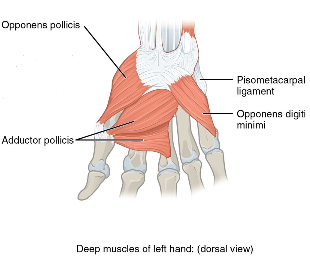
Delving into the hand’s deeper layers reveals its intricate design. The image presents the deep muscles of the left hand from a dorsal perspective, with each muscle clearly labeled for study.
- Lumbricalis muscles: Originating from the flexor digitorum profundus tendons, they flex the metacarpophalangeal joints and extend the interphalangeal joints.
- Dorsal interossei muscles: Arising from the metacarpal bones, they abduct the fingers away from the middle finger.
- Extensor indicis: Stemming from the ulna, it extends the index finger for individual digit control.
- Extensor pollicis longus: Originating from the ulna, it extends the distal phalanx of the thumb.
- Extensor pollicis brevis: Arising from the radius, it extends and abducts the proximal phalanx of the thumb.
- Abductor pollicis longus: Stemming from the radius and ulna, it abducts and extends the thumb.
Anatomical Overview
Exploring the deep dorsal structure highlights its specialized arrangement. The lumbricalis muscles, dorsal interossei muscles, extensor indicis, extensor pollicis longus, extensor pollicis brevis, and abductor pollicis longus form the deep layer, each contributing to the hand’s range of motion.
- The lumbricalis muscles enhance finger coordination across multiple joints.
- The dorsal interossei muscles facilitate finger abduction for hand spreading.
- The extensor indicis provides independent extension of the index finger.
- The extensor pollicis longus and brevis manage thumb extension and stability.
- The abductor pollicis longus supports thumb abduction and extension.
Functional Roles of Deep Hand Muscles
Understanding the functional contributions emphasizes their role in dexterity. These muscles work together to perform precise movements, from finger abduction to thumb extension, relying on their deep placement.
- The lumbricalis muscles coordinate flexion at the metacarpophalangeal joints and extension at the interphalangeal joints.
- The dorsal interossei muscles abduct the fingers, essential for hand widening.
- The extensor indicis extends the index finger, key for pointing or writing.
- The extensor pollicis longus extends the thumb’s distal phalanx, aiding precision tasks.
- The extensor pollicis brevis extends and abducts the thumb’s proximal phalanx, enhancing grip.
- The abductor pollicis longus abducts and extends the thumb, vital for grasping.
Clinical Significance
Investigating the clinical implications underscores their practical importance. Injuries or dysfunctions in these deep muscles can impair hand function, requiring specific rehabilitation strategies.
- Strain in the lumbricalis muscles can affect finger coordination, often treated with therapy.
- The dorsal interossei muscles injury may limit finger abduction, needing exercises.
- The extensor indicis dysfunction can impair index finger extension, managed with rehabilitation.
- The extensor pollicis longus damage may reduce thumb extension, requiring targeted care.
- The extensor pollicis brevis strain can weaken thumb movement, treated with rest and therapy.
- The abductor pollicis longus injury may hinder thumb abduction, necessitating intervention.
Conclusion
The study of deep muscles of the left hand from a dorsal view reveals a sophisticated network of anatomy and function. The lumbricalis muscles, dorsal interossei muscles, extensor indicis, extensor pollicis longus, extensor pollicis brevis, and abductor pollicis longus each play a unique role in providing fine motor control and stability to the hand. This understanding not only deepens appreciation of the hand’s deeper structure but also supports effective management of related injuries, enhancing overall hand health and functionality.

