The hand is a remarkable structure, relying on its intrinsic muscles to provide the fine motor control essential for daily tasks, with all origins and insertions located within the hand itself. This article delves into the superficial muscles of the left hand as depicted in a palmar view, highlighting their roles in flexing, extending, abducting, and adducting the distal segments of the fingers and thumb. The detailed illustration serves as a key resource for understanding hand anatomy and its functional significance in clinical contexts.
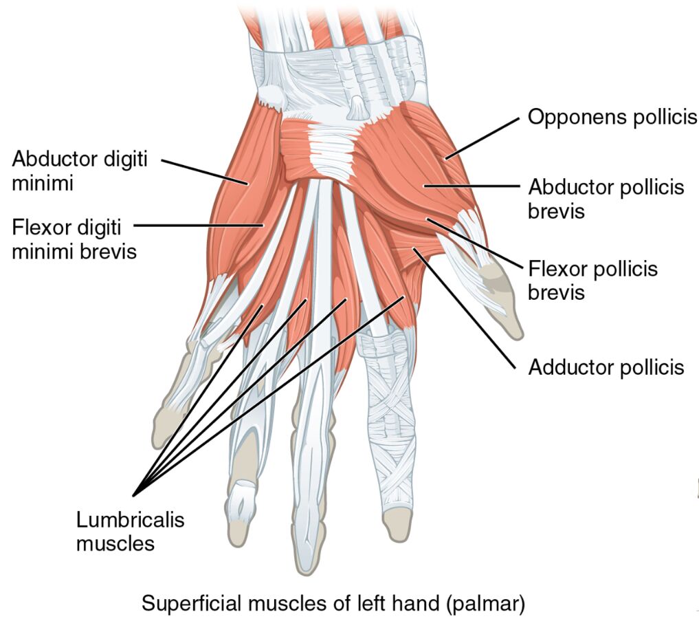
Unveiling the hand’s superficial layer offers insight into its mobility. The image showcases the superficial muscles of the left hand from a palmar perspective, with each muscle distinctly labeled.
- Abductor digiti minimi: Originating from the pisiform bone, it abducts the little finger to widen the hand.
- Flexor digiti minimi brevis: Arising from the hamate bone, it flexes the proximal phalanx of the little finger.
- Lumbricalis muscles: Originating from the flexor digitorum profundus tendons, they flex the metacarpophalangeal joints and extend the interphalangeal joints.
- Opponens pollicis: Stemming from the trapezium and flexor retinaculum, it opposes the thumb for grasping actions.
- Abductor pollicis brevis: Arising from the scaphoid and trapezium, it abducts the thumb away from the hand.
- Flexor pollicis brevis: Originating from the flexor retinaculum and trapezium, it flexes the proximal phalanx of the thumb.
- Adductor pollicis: Originating from the capitate and metacarpal bones, it adducts the thumb toward the fingers.
Anatomical Overview
Delving into the superficial muscular structure reveals a specialized design. The abductor digiti minimi, flexor digiti minimi brevis, lumbricalis muscles, opponens pollicis, abductor pollicis brevis, flexor pollicis brevis, and adductor pollicis form the palmar superficial layer, each contributing to distinct hand movements.
- The abductor digiti minimi and flexor digiti minimi brevis control the little finger’s range of motion.
- The lumbricalis muscles enhance finger flexibility and extension across multiple joints.
- The opponens pollicis, abductor pollicis brevis, and flexor pollicis brevis manage thumb opposition and flexion.
- The adductor pollicis facilitates thumb adduction, strengthening grip.
Functional Roles of Superficial Hand Muscles
Understanding the functional contributions underscores their importance in dexterity. These muscles work in harmony to execute precise movements, from thumb opposition to finger flexion, relying on their palmar positioning.
- The abductor digiti minimi spreads the little finger, aiding in hand expansion.
- The flexor digiti minimi brevis flexes the little finger, supporting grip stability.
- The lumbricalis muscles coordinate flexion at the metacarpophalangeal joints and extension at the interphalangeal joints.
- The opponens pollicis enables thumb opposition, essential for grasping objects.
- The abductor pollicis brevis abducts the thumb, enhancing hand versatility.
- The flexor pollicis brevis flexes the thumb, crucial for holding items.
- The adductor pollicis adducts the thumb, reinforcing grip strength.
Clinical Significance
Investigating the clinical implications highlights their practical relevance. Injuries or dysfunctions in these superficial muscles can impair hand function, necessitating targeted rehabilitation approaches.
- Strain in the abductor digiti minimi can limit little finger abduction, often treated with therapy.
- The flexor digiti minimi brevis injury may weaken little finger flexion, requiring exercises.
- The lumbricalis muscles damage can affect finger coordination, managed with rehabilitation.
- The opponens pollicis dysfunction can reduce thumb opposition, treated with physical therapy.
- The abductor pollicis brevis injury may impair thumb abduction, needing care.
- The flexor pollicis brevis strain can weaken thumb flexion, managed with rest.
- The adductor pollicis damage may hinder thumb adduction, necessitating intervention.
Conclusion
The exploration of superficial muscles of the left hand in a palmar view reveals a sophisticated network of anatomy and function. The abductor digiti minimi, flexor digiti minimi brevis, lumbricalis muscles, opponens pollicis, abductor pollicis brevis, flexor pollicis brevis, and adductor pollicis each play a unique role in providing fine motor control and stability to the hand. This understanding not only deepens appreciation of the hand’s superficial structure but also supports effective management of related injuries, enhancing overall hand health and functionality.

