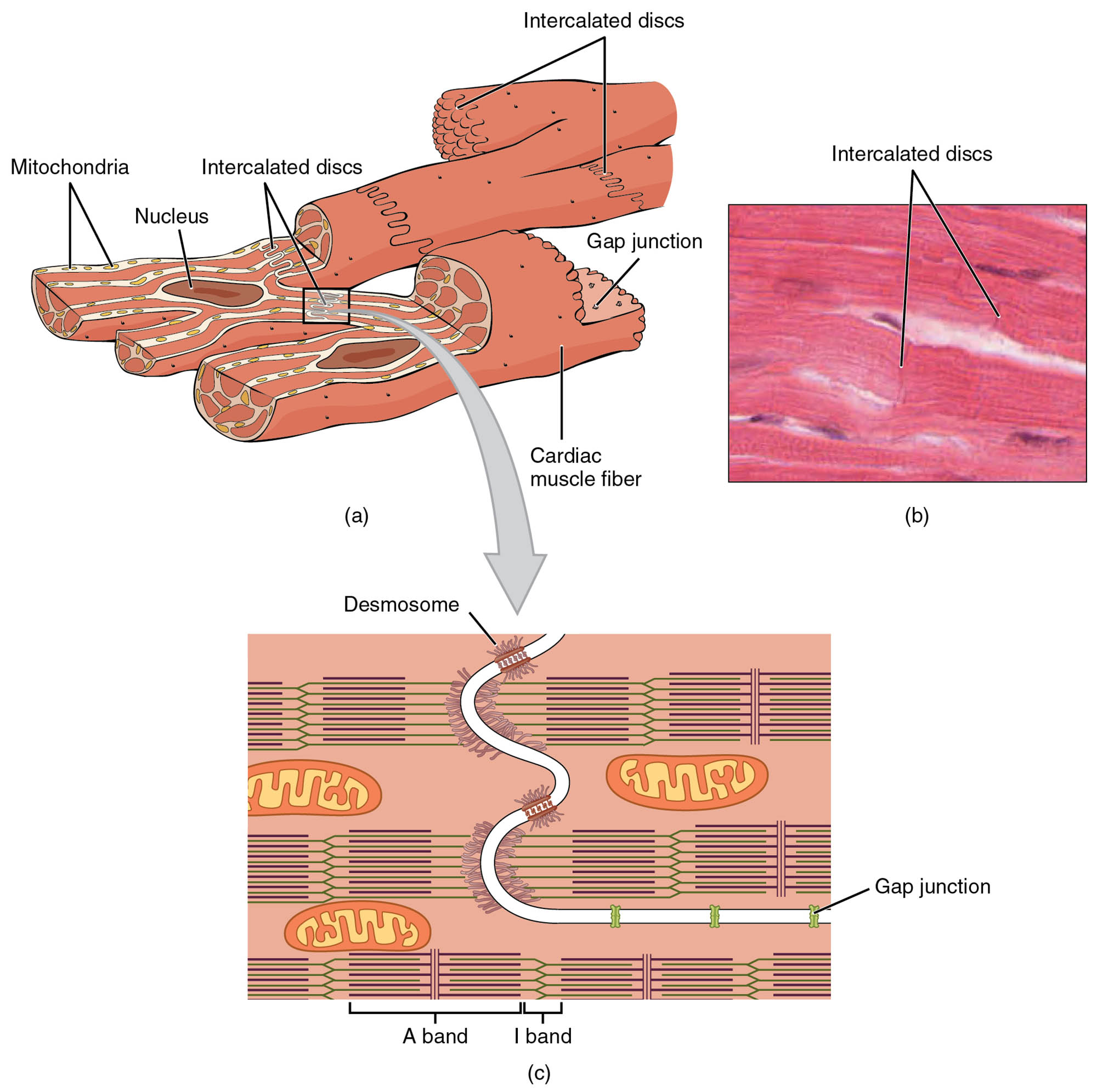
The cardiac muscle cell is a cornerstone of the heart’s ability to pump blood, featuring a unique microscopic structure that supports its continuous function. This diagram and photomicrograph illustrate the intricate details of myofibrils, sarcomeres, T tubules, mitochondria, intercalated discs, nuclei, desmosomes, and gap junctions, providing a window into the cellular architecture that drives cardiac performance. Exploring these components offers valuable insights into the heart’s remarkable endurance and efficiency.
Labelled Parts Explanation
- Myofibrils The myofibrils are elongated thread-like structures within cardiac muscle cells, composed of repeating units called sarcomeres. They are responsible for the contractile force that enables the heart to pump blood effectively.
- Sarcomeres The sarcomeres are the basic contractile units of myofibrils, containing actin and myosin filaments that slide past each other during contraction. Their organized arrangement contributes to the heart’s rhythmic beating.
- T tubules The T tubules are invaginations of the sarcolemma that penetrate deep into the cell, transmitting action potentials to the interior. This ensures synchronized contraction across the cardiac muscle cell.
- Mitochondria The mitochondria are numerous within cardiac muscle cells, producing ATP to fuel continuous contraction. Their high density reflects the heart’s constant energy demands.
- Intercalated discs The intercalated discs are specialized junctions between adjacent cardiac muscle cells, facilitating electrical and mechanical coupling. They contain desmosomes and gap junctions to maintain heart tissue integrity.
- Nuclei The nuclei are centrally located within cardiac muscle cells, containing the genetic material that directs cellular function. Each cell typically has one or two nuclei, supporting the cell’s metabolic activity.
- Desmosomes The desmosomes are strong adhesion points within intercalated discs, anchoring cardiac muscle cells together. They prevent separation of cells during the heart’s forceful contractions.
- Gap junctions The gap junctions are channels within intercalated discs that allow rapid electrical impulse transmission between cells. This connectivity ensures coordinated contraction across the heart.
Anatomical Overview of Cardiac Muscle Cells
The cardiac muscle cell’s structure is tailored for its relentless pumping role. This diagram and photomicrograph reveal the cellular components that sustain heart function.
- The myofibrils and sarcomeres form the contractile machinery, driving blood movement.
- The T tubules and mitochondria support rapid signaling and energy production, respectively.
- The intercalated discs, with desmosomes and gap junctions, link cells for unified action.
- The nuclei oversee cellular maintenance, ensuring long-term viability.
This microscopic view highlights the heart’s adaptation to continuous operation.
Structure and Function of Myofibrils and Sarcomeres
Myofibrils and sarcomeres are the heart’s contractile foundation. Their organization enables efficient muscle action.
- The myofibrils run parallel within the cell, packed with sarcomeres for contraction.
- The sarcomeres’ actin and myosin filaments slide during systole, shortening the muscle.
- This process generates the force needed to pump blood through the circulatory system.
- The striped appearance under a microscope reflects their precise alignment.
These structures are essential for the heart’s rhythmic performance.
Role of T Tubules and Mitochondria
T tubules and mitochondria are critical for cardiac cell function. They ensure rapid response and energy supply.
- The T tubules carry electrical impulses from the sarcolemma, triggering calcium release.
- The mitochondria produce ATP via oxidative phosphorylation, supporting constant contraction.
- Their abundance provides the energy for the heart’s 24/7 operation.
- This synergy enhances the cell’s ability to respond to demand.
These components underpin the heart’s endurance.
Significance of Intercalated Discs
Intercalated discs are unique to cardiac muscle, ensuring cell coordination. Their structure supports heart unity.
- The intercalated discs connect adjacent cells, containing desmosomes for strength.
- The gap junctions within these discs allow ion flow, enabling synchronized contraction.
- This connectivity is vital for the heart to function as a single unit.
- The discs’ design prevents tearing under mechanical stress.
This feature is key to the heart’s cohesive action.
Microscopic Features in Photomicrographs
The photomicrograph reveals cardiac muscle’s cellular details. These visual elements provide structural insight.
- The nuclei appear as dark, central spots, indicating active cellular metabolism.
- The intercalated discs are visible as dark lines, showing cell junctions at 1600x magnification.
- The striated pattern reflects the sarcomeres’ alignment within myofibrils.
- This high-resolution view aids in studying cellular anatomy.
The image enhances understanding of cardiac tissue organization.
Physiological Importance of Cardiac Muscle Structure
The cardiac muscle cell’s anatomy supports its physiological demands. This structure optimizes heart performance.
- The T tubules ensure uniform calcium distribution for contraction.
- The mitochondria’s high density meets the energy needs of continuous pumping.
- The intercalated discs’ desmosomes and gap junctions maintain electrical and mechanical sync.
- This design allows the heart to adapt to varying workloads.
The cell’s architecture is critical for sustaining circulation.
Clinical Relevance of Cardiac Muscle Cells
Understanding cardiac muscle cell structure aids in diagnosing heart conditions. These components are key clinical markers.
- Disruption of intercalated discs’ gap junctions can lead to arrhythmias.
- Reduced mitochondria function may contribute to cardiomyopathy.
- Damage to sarcomeres is seen in myocardial infarction.
- Imaging and biopsy assess these structures for disease management.
This knowledge supports effective cardiac care and treatment.
Conclusion
The cardiac muscle cell anatomical structure and microscopic view provide a detailed exploration of the cellular elements that power the heart. By examining the myofibrils, sarcomeres, T tubules, mitochondria, intercalated discs, nuclei, desmosomes, and gap junctions, one gains insight into the heart’s ability to contract efficiently and endure tirelessly. This understanding serves as a foundation for studying cardiovascular physiology and addressing related health issues, encouraging further exploration of the heart’s intricate cellular design and its critical role in sustaining life.

