The heart’s rhythmic beating is governed by a precise electrical conduction system, depicted step-by-step in this informative diagram. This image traces the process from the sinoatrial (SA) node initiating an action potential to the ventricular contractile fibers contracting, including key stages like the atrioventricular (AV) node delay and the role of the moderator band. Delving into this diagram offers a comprehensive view of how electrical impulses coordinate the heart’s pumping action to sustain circulation.
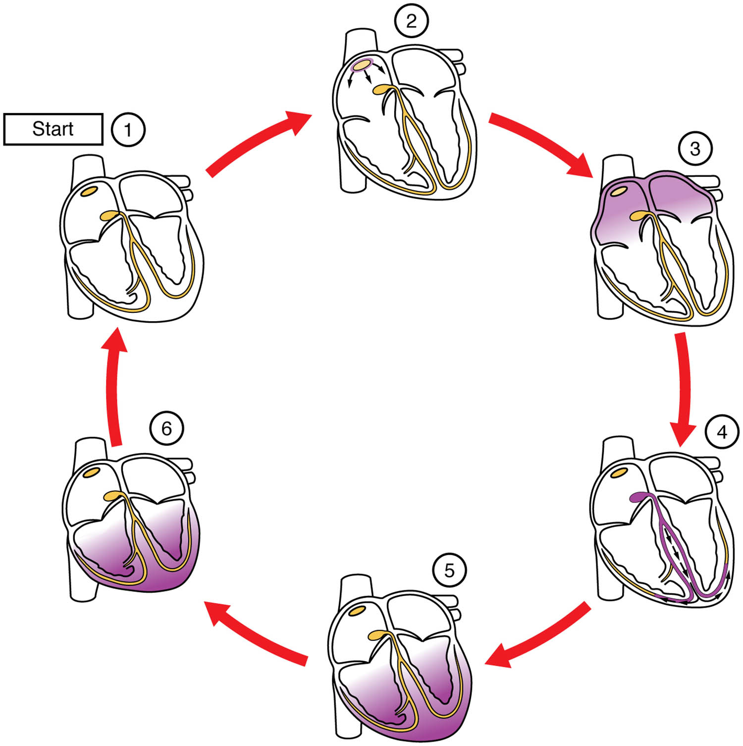
Labelled Parts Explanation
- Sinoatrial (SA) node at rest The sinoatrial (SA) node at rest is the heart’s natural pacemaker, located in the right atrium, preparing to initiate the cardiac cycle. It remains quiescent until triggered to generate an electrical impulse.
- Sinoatrial (SA) node initiates action potential The sinoatrial (SA) node initiates action potential by depolarizing, sending an electrical wave across the atria to trigger contraction. This marks the beginning of the heartbeat and sets the rhythm.
- Atrioventricular (AV) node delay The atrioventricular (AV) node delay slows the impulse for approximately 100 ms, allowing the atria to fully contract and empty blood into the ventricles. This pause ensures proper timing in the cardiac cycle.
- Atrioventricular (AV) bundle and bundle branches The atrioventricular (AV) bundle and bundle branches conduct the impulse from the AV node through the interventricular septum to the ventricles. They divide into right and left branches to distribute the signal efficiently.
- Purkinje fibers The Purkinje fibers spread the electrical impulse rapidly across the ventricular myocardium, ensuring synchronized contraction. They enable the ventricles to pump blood effectively with each beat.
- Moderator band to right papillary muscle The moderator band to right papillary muscle carries the impulse to the right ventricle’s papillary muscle, aiding valve function. It provides a direct conduction path to support right ventricular contraction.
- Ventricular contractile fibers The ventricular contractile fibers are the muscle cells that contract in response to the Purkinje fiber impulse, ejecting blood into the pulmonary artery and aorta. Their activation marks the final stage of ventricular systole.
- Ventricular contraction begins The ventricular contraction begins as the contractile fibers activate, reducing the ventricular volume to pump blood out. This phase completes the cardiac cycle’s pumping action.
Anatomical Overview of the Cardiac Conduction System
The cardiac conduction system orchestrates the heart’s rhythmic contractions. This diagram illustrates the sequential activation that drives blood flow.
- The sinoatrial (SA) node at rest prepares to initiate the cycle, while the sinoatrial (SA) node initiates action potential starts the process.
- The atrioventricular (AV) node delay ensures atrial emptying before ventricular action.
- The atrioventricular (AV) bundle and bundle branches and Purkinje fibers distribute the impulse to the ventricles.
- The moderator band to right papillary muscle and ventricular contractile fibers complete the contraction phase.
This pathway maintains the heart’s ability to pump continuously.
Initiation and Spread of the Action Potential
The action potential’s initiation sets the cardiac cycle in motion. This stage is critical for rhythm establishment.
- The sinoatrial (SA) node initiates action potential generates the electrical signal at 60-100 beats per minute.
- The impulse sweeps across the atria, causing them to contract and push blood into the ventricles.
- The sinoatrial (SA) node at rest phase allows recovery before the next cycle.
- This process is regulated by autonomic nervous system inputs.
The initial signal is the foundation of cardiac rhythm.
Role of the Atrioventricular Node and Delay
The atrioventricular node introduces a vital delay in the conduction process. This pause optimizes blood flow.
- The atrioventricular (AV) node delay allows the atria to complete systole, filling the ventricles.
- The delay, lasting about 100 ms, is due to slower conduction properties of the AV node.
- This timing prevents overlap between atrial and ventricular contractions.
- The node acts as a gatekeeper for ventricular activation.
This mechanism ensures efficient cardiac output.
Distribution via Bundle Branches and Purkinje Fibers
The bundle branches and Purkinje fibers ensure rapid ventricular activation. Their role is key to synchronized pumping.
- The atrioventricular (AV) bundle and bundle branches carry the impulse to the right and left ventricles.
- The Purkinje fibers distribute the signal quickly, triggering ventricular contractile fibers.
- Their fast conduction velocity maximizes ventricular ejection.
- The moderator band to right papillary muscle supports right ventricular coordination.
This network enhances the heart’s pumping efficiency.
Completion of Ventricular Contraction
Ventricular contraction marks the end of the electrical conduction cycle. This phase drives blood circulation.
- The ventricular contractile fibers contract in response to the Purkinje fiber impulse.
- The ventricular contraction begins phase ejects blood into the pulmonary and systemic circuits.
- The moderator band to right papillary muscle ensures valve stability during this process.
- This action completes the systolic phase of the cardiac cycle.
The contraction is essential for maintaining circulation.
Physiological Importance of the Conduction System
The conduction system’s design supports the heart’s continuous operation. Its structure optimizes performance.
- The sinoatrial (SA) node initiates action potential adapts to physiological demands.
- The atrioventricular (AV) node delay ensures proper filling and ejection timing.
- The Purkinje fibers and ventricular contractile fibers enable powerful, synchronized beats.
- This system maintains a steady heart rate and rhythm.
The coordination is vital for cardiovascular health.
Clinical Relevance of the Conduction System
Understanding the conduction system aids in managing cardiac arrhythmias. These components are key clinical targets.
- Failure of the sinoatrial (SA) node initiates action potential can lead to bradycardia or tachycardia.
- Blockage at the atrioventricular (AV) node delay causes AV block, disrupting rhythm.
- Damage to the Purkinje fibers may result in ventricular fibrillation.
- Interventions like pacemakers target these structures for treatment.
This knowledge supports effective cardiac care.
Conclusion
The cardiac conduction diagram provides a detailed roadmap of the electrical pathway that governs the heart’s rhythm and pumping action. By tracing the journey from the sinoatrial (SA) node at rest to the ventricular contraction begins, including the atrioventricular (AV) node delay and Purkinje fibers, one gains insight into the heart’s coordinated function. This understanding serves as a foundation for studying cardiovascular physiology and addressing related health challenges, encouraging further exploration of the heart’s intricate electrical design and its critical role in sustaining life.

